Medicine and Health
Post-operative fracture risk assessment following tumor curettage in the distal femur: a hybrid in vitro and in silico biomechanical approach
A. Ghouchani, G. Rouhi, et al.
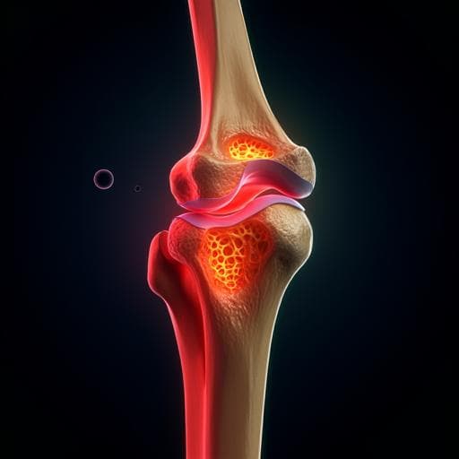
This groundbreaking study by Azadeh Ghouchani, Gholamreza Rouhi, and Mohammad Hosein Ebrahimzadeh introduces a biomechanical tool to evaluate post-operative fracture risk after distal femur tumor curettage. With the help of finite element models and quantitative CT imaging, the research uncovers critical insights into bone strength and reconstruction vulnerabilities.
Related Publications
Explore these studies to deepen your understanding of the subject.


