
Medicine and Health
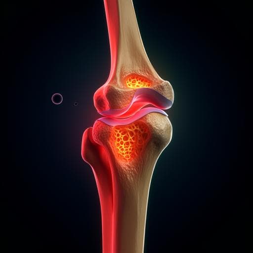
Post-operative fracture risk assessment following tumor curettage in the distal femur: a hybrid in vitro and in silico biomechanical approach
A. Ghouchani, G. Rouhi, et al.
This groundbreaking study by Azadeh Ghouchani, Gholamreza Rouhi, and Mohammad Hosein Ebrahimzadeh introduces a biomechanical tool to evaluate post-operative fracture risk after distal femur tumor curettage. With the help of finite element models and quantitative CT imaging, the research uncovers critical insights into bone strength and reconstruction vulnerabilities.
~3 min • Beginner • English
Introduction
Benign tumours such as giant cell tumour (GCT) often occur around the knee and can create lytic lesions that weaken load-bearing bones like the distal femur, leading to a substantial risk of post-operative fracture after curettage. Existing clinical criteria for pathologic fracture risk (e.g., Mirels’ and Harrington’s) have low specificity and are not tailored to post-operative risk following tumour removal. There is no quantitative, validated criterion to determine which defects require cement augmentation or which fixation devices best reduce post-operative fracture risk. The study aims to develop and validate a QCT-based, non-linear finite element approach to predict distal femoral strength after simulated curettage, identify critical defect size and location thresholds for increased fracture risk, and investigate mechanics at the bone-cement interface to inform augmentation strategies.
Literature Review
Prior QCT-based FE studies have predicted strength in proximal femur and vertebrae with promising accuracy. In tumour contexts, FE and rigidity analyses have been used to assess fracture risk and reconstruction strategies, but many prior works: (1) focused on pathologic (pre-operative) rather than post-operative risk; (2) used linear or homogeneous models, reducing accuracy; or (3) lacked validation with experiments or clinical data. Retrospective CT-based FE work suggested many prophylactic stabilizations flagged by Mirels’ might be unnecessary. Some studies reported acceptable outcomes without filling defects, while others advocated PMMA and augmentation. A retrospective series indicated post-curettage fracture risk increases with larger defects and certain locations. Device selection for augmentation remains controversial, partly due to limited understanding of bone–cement interface mechanics; previous in vitro or FE studies often neglected interface behavior or lacked validation.
Methodology
Design: Hybrid experimental-computational study using paired human cadaveric distal femora to validate subject-specific QCT-based non-linear heterogeneous FE models, followed by in silico parametric analyses of defect size and location and assessment of bone–cement interface mechanics.
Specimens and imaging: Seven pairs (n=14) of healthy human cadaveric distal femora (4 male, 3 female; mean age 37, range 32–64). One femur per pair received a simulated tumour curettage defect in the epiphysis with a smoothed interior surface and PMMA cement infill; the contralateral femur remained intact as control. QCT scans performed immersed in water using a clinical scanner (Siemens Somatom 64; 140 kV, 80 mAs; in-plane 0.5×0.5 mm; 1 mm slice thickness) with a calibration phantom to convert HU to ash density.
Mechanical testing: Specimens mounted with the diaphysis aligned vertically (z-axis). The proximal end was embedded to restrain all DOF. Compression load applied via a 25 mm diameter actuator at 1 mm/min to the medial condyle, parallel to the shaft axis. Reaction force–displacement recorded; maximum force taken as fracture load (FExp).
FE model construction: Voxel-based meshes generated from QCT (Simpleware ScanIP/ScanFE) with each voxel converted to an 8-node brick element. Heterogeneous isotropic material properties assigned from ash density using Keyak et al. relationships for distal femur: E=14900·ρash^1.80 (MPa); yield stress S=102·ρash^1.80 (MPa); quad-linear constitutive law with elastic phase, perfect plastic phase (plastic strain εAB; trabecular: εAB=0.00189+0.0241·ρash; cortical: εAB=0.0184−0.0100·ρash), softening with plastic modulus Ep (trabecular: Ep=−20800·ρash^1.45 MPa; cortical: Ep=−1000 MPa), followed by a second perfect plastic phase; minimum stress σmin=43.1·ρash^1.81 (MPa). PMMA cement assigned homogeneous properties E=2 GPa, ν=0.28, strength=40 MPa.
Boundary/loading conditions: Proximal end constrained; compression applied on nodes within a 25 mm diameter circular footprint on the medial condyle (as in the test setup). For bilateral loading simulations (defect size/location study), load distributed 60% medial and 40% lateral.
Failure criterion and solver: Non-linear analyses performed in ABAQUS (v6.13-4). Distortion energy (von Mises) failure theory used to predict failure load. Force–displacement curves computed; maximum force defined as FE-predicted fracture load (FFE).
Model validation and tuning: Models split into Tuning Group (TG, n=5) and Evaluation Group (EG, n=9). In TG, density-based material parameters (E, S, Ep) were uniformly reduced in 1% steps using custom MATLAB until mean prediction error μ=FFE−FExp was not different from zero via paired t-test, partially compensating for isotropy and other systematic modeling errors. The optimal reduction (17%) was then applied to EG for independent validation. Statistics: paired t-tests, linear regression, Pearson correlation (SPSS v16; α=0.05).
Defect size and location analyses: Starting from a validated defective model (medial condyle defect DV=36 cc), the defect was dilated in all directions to generate DV=36, 44, 52, 65, 74, 83, 91, 98 cc. Epiphyseal volume (EPV) defined from the distal end to diaphyseal onset; DV/EPV (%) computed. For each DV, FFE compared to intact. One-sample t-tests compared mean FFE of groups (<65 cc vs >65 cc) to intact. Larger-than-critical defects (76–98 cc) mirrored to medial and lateral compartments to compare location effects; invasion of the contralateral condyle was noted when DV/EPV exceeded ~47%.
Bone–cement interface (BCI) mechanics: Contact and damage initiation at the BCI evaluated through FE post-processing. Regions reaching mixed-mode damage initiation threshold (τ0) identified (maximum stress criterion). Contact status mapped to detect sticking, slipping, and debonded areas to locate vulnerable regions for reconstruction failure.
Key Findings
- Validation: After a 17% reduction in density-based material parameters during tuning, TG achieved μ=375 N (P>0.05). Applying the same reduction to EG yielded μ=129 N (P>0.05), indicating no significant difference between FFE and FExp. Across all 14 specimens, paired t-test showed no significant difference (P=0.174). Pearson correlation r=0.97 (P<0.001). Linear regression: FFE=1.12·FExp−0.570, R²=0.95 (P<0.001). 95% CI of FFE−FExp had a maximum of 542 N.
- Critical defect size: Minimal strength reduction (<5%) until DV≈65 cc, corresponding to DV/EPV≈35%. Mean FFE for defects <65 cc was 4820 N vs intact 4908 N (no significant difference, P>0.05). Mean FFE for defects >65 cc was 3629 N, significantly lower than intact (P<0.05). When DV reached 35% EPV, defective bone retained ~95% of intact strength; at 45% EPV, strength dropped to ~70% of intact.
- Sensitivity to DV: For DV below critical, strength decreased ~0.06% per 1 cc increase; above critical, ~1.14% per 1 cc increase.
- Location effects: For DV in the 39–46% EPV range, lateral defects showed less reduction in strength than medial defects of similar size/shape; lateral defects could be on average ~4% larger (DV/EPV) to produce similar strength reduction as medial. Above ~50% EPV, medial–lateral differences diminished. When DV/EPV exceeded ~47% and the defect invaded the contralateral condyle, a sharper strength reduction occurred.
- Bone–cement interface (BCI): Damage initiation concentrated at the proximal end of the cortical window. Contact analysis revealed a larger cement debonding area at the most interior wall of the interface, particularly where the defect extended toward the intercondylar region.
Discussion
The study addressed the lack of a quantitative, validated criterion for post-operative fracture risk after curettage by developing and validating a patient-specific, non-linear QCT-based FE framework. Strong agreement with in vitro failure loads demonstrates its clinical potential. A key result is a volumetric, site-specific threshold: defects exceeding ~65 cc or ~35% of epiphyseal volume lead to substantial strength loss and heightened fracture risk, supporting retrospective clinical observations that larger defects substantially elevate post-operative fracture risk. The importance of defect location emerged: laterally located epiphyseal defects tend to be biomechanically safer than medial defects of equivalent size, a factor omitted in existing clinical scoring systems. Involvement of the contralateral condyle is an additional risk amplifier.
Analysis of the bone–cement interface identified vulnerable zones for reconstruction failure—the proximal edge of the cortical window and the most interior interface wall with a propensity for debonding—providing biomechanical rationale for selecting augmentation strategies that reinforce these regions. These insights align with prior experimental observations and suggest that plate constructs, which bridge the cortical window and allow screw purchase across the interior wall, may better mitigate failure risk than intramedullary pins that do not address these vulnerable areas. Overall, the findings support integrating 3D volumetric defect metrics and anatomical location into post-operative risk assessment and planning of cement augmentation.
Conclusion
A validated QCT-based non-linear FE approach accurately predicted fracture loads of intact and post-curettage distal femora and identified critical determinants of post-operative fracture risk. Bone strength showed a marked decline once defect size exceeded ~35% of the epiphyseal volume (~65 cc), with laterally located defects carrying lower risk than medial defects of the same size. Vulnerable regions for reconstruction failure were the proximal edge of the cortical window and the most interior bone–cement interface wall. Future work should develop comprehensive analytical criteria incorporating 3D defect size, shape, and location, contralateral condyle involvement, and patient-specific factors (bone quality, activity, body weight), and further refine implant selection and placement strategies targeting the identified high-risk interface regions.
Limitations
- Bone modeled as isotropic with density-based properties originally derived for the longitudinal direction; anisotropy and multiaxial differences (tension vs compression) were not explicitly modeled, partially compensated by material parameter tuning.
- Only axial compressive loading was evaluated; torsional and bending loads, which also contribute to failures, were not analyzed.
- No standardized defect shape was enforced; while volumetric dilation captured size effects, shape variability remains a factor.
- Critical defect size was determined primarily for medially located defects; lateral critical thresholds require further study.
- Augmentation device recommendations were inferred from BCI analyses without explicitly modeling implants and their contacts; full implant–bone–cement models are needed to account for load transfer and fixation mechanics.
- Potential additional systematic errors (e.g., boundary conditions, mesh size effects) may persist despite tuning.
Related Publications
Explore these studies to deepen your understanding of the subject.