Medicine and HealthBone Research
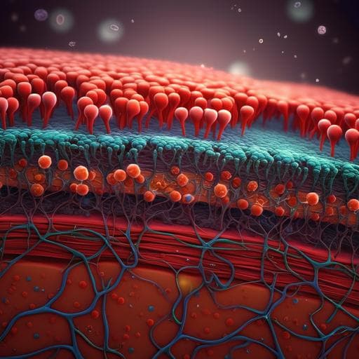
Construction of developmentally inspired periosteum-like tissue for bone regeneration
K. Dai, S. Deng, et al.
This groundbreaking research by Kai Dai and collaborators focuses on innovative periosteum-like tissue engineering using BMP-2-loaded scaffolds. The creation of tissue that mimics natural bone development promises significant advancements in regenerative medicine, especially for older populations. Discover how the addition of chondroitin sulfate enhances bone regenerative capabilities!
Related Publications
Explore these studies to deepen your understanding
Adjacent work that informs or extends this paper's methodology and findings.

Food Science and Technology
Formation of contractile 3D bovine muscle tissue for construction of millimetre-thick cultured steak
M. Furuhashi, Y. Morimoto, et al.

Medicine and Health
Sequential laxative-probiotic usage for treatment of irritable bowel syndrome: a novel method inspired by mathematical modelling of the microbiome
M. Li, R. Xu, et al.
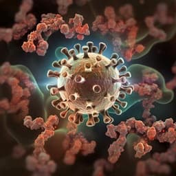
Medicine and Health
Robust induction of functional humoral response by a plant-derived Coronavirus-like particle vaccine candidate for COVID-19
P. Kaplonek, D. Cizmeci, et al.
Food Science and Technology
Generation of three-dimensional meat-like tissue from stable pig epiblast stem cells
G. Zhu, D. Gao, et al.


