Medicine and Health
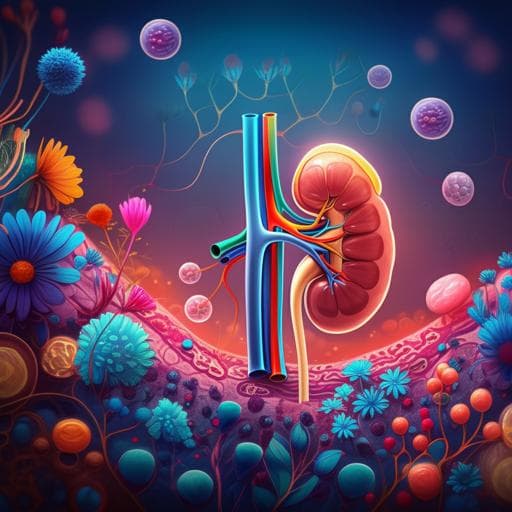
Vitamin D is involved in the effects of the intestinal flora and its related metabolite TMAO on perirenal fat and kidneys in mice with DKD
X. Zheng, Y. Huang, et al.
This study reveals how vitamin D plays a crucial role in improving gut health, reducing kidney damage, and influencing perirenal adipose tissue in diabetic kidney disease. Conducted by a team from Shanghai Fifth People's Hospital and Fudan University, the findings suggest exciting connections between vitamin D and kidney protection.
~3 min • Beginner • English
Introduction
The gut microbiota functions as a key "organ" influencing host immunity, digestion, endocrine regulation, neural signaling, drug metabolism, and nutrient absorption. Microbial metabolites act on receptors in liver, intestine, brown and white adipose tissue, and CNS, affecting nutrient synthesis, motility, and electrolyte balance. Dietary choline is metabolized by gut flora to TMA and oxidized in liver to TMAO, implicated in atherosclerosis, hepatic steatosis, and is associated with vitamin D deficiency and NAFLD. TMAO has also been linked to diabetes, CKD, and colorectal cancer, and is associated with renal fibrosis and impairment; however, mechanistic links with DKD remain unclear. Vitamin D/VDR signaling is renoprotective and interacts with gut microbes, favoring probiotic colonization and inhibiting NF-κB activation, with VD deficiency linked to alterations in intestinal and renal VDR expression. PRAT, anatomically adjacent to kidneys and adrenal glands, contains brown and beige adipocytes with endocrine functions; its expansion and inflammation may contribute to renal injury. TMAO production has been associated with inhibition of beige/WAT activities and is an attractive target in obesity-related diseases. Markers of WAT browning include UCP-1 upregulation and mitochondrial biogenesis. The study aims to explore the effects of administering different concentrations of 1,25-(OH)2D3 on the TLR4/NF-κB pathway, intestinal flora, and kidney tissue in DKD mice; to evaluate the relationship between serum TMAO and urine ACR; and to determine whether TMAO influences PRAT metabolism and whether vitamin D modulates TMAO effects via VDR in PRAT and kidney, providing insights for DKD prevention and treatment.
Literature Review
Prior work links high circulating TMAO to cardiovascular disease, NAFLD, diabetes, CKD, and increased mortality in CKD. Gut microbial taxa (e.g., Enterobacteriaceae, Escherichia, Desulfovibrio, Clostridium, Enterococcus, Streptococcus, Actinomycetes) encode enzymes for TMA production from choline/carnitine. VD/VDR signaling is renoprotective, inhibits NF-κB in renal cells, and modulates gut microbiota favoring probiotics during inflammation, improving intestinal barrier integrity. VD deficiency correlates with CKD risk and poorer outcomes, while supplementation may improve survival. Observational studies report inverse associations between VD levels and TMAO, particularly in obesity and NAFLD, and VD supplementation can reduce fasting TMAO and improve choline metabolism. PRAT expansion is linked to renal dysfunction, and beige/brown adipose thermogenesis influences metabolic health. These findings provide context for investigating VD–microbiota–TMAO interactions affecting PRAT and kidney in DKD.
Methodology
Animals: Sixty male KKay (type 2 diabetes) mice and 10 C57BL/6 controls (8 weeks old; 18–22 g) were housed in SPF conditions. KKay mice were fed a high-sugar, high-fat, high-cholesterol diet (formulation detailed) to 20 weeks. Experimental groups: Of 56 KKay mice, DM group (n=10; 14 weeks), DKD group (n=46; 20 weeks), plus C57BL/6 controls (n=10; 20 weeks). DKD mice were randomized to placebo (n=10), low-dose VD (LVD; 0.3 µg/kg/d; n=10), medium-dose VD (MVD; 1.4 µg/kg/d; n=16), high-dose VD (HVD; 2.5 µg/kg/d; n=10) with daily 1,25-(OH)2D3 or saline injections from 18 to 20 weeks. Six DKD mice received 0.12% dietary TMAO concurrently with MVD. Endpoints: feces, urine, serum, PRAT, and kidneys collected at 20 weeks. Measurements: Body weight, tail vein blood glucose; urine in metabolic cages for protein excretion, microalbumin, creatinine; UACR calculated; assays on AU5821 analyzer. Serum TNF-α measured by ELISA; serum and urine TMAO by LC-MS (UPLC BEH HILIC column). Urinary KIM-1 by ELISA. Molecular analyses: qPCR for TLR4, NF-κB, PGC1α, UCP-1 (2^-ΔΔCt; primer sequences provided). 16S rRNA V3–V4 amplicon sequencing (338F/806R) on NovaSeq; alpha/beta diversity and taxonomic composition analyses; intergroup differences by Metastats. Histology: Kidney HE, PAS, Masson staining; PRAT HE and Oil Red O. Immunofluorescence: podocin, UCP-1; IHC: VDR, PGC1α, UCP-1 in kidney and PRAT. Electron microscopy: renal and PRAT ultrastructure; GBM thickness measured across fields. VDR knockdown: Lentiviral shRNA vectors targeting VDR (Gene ID 22337) constructed; titers tested in 293T cells; best-performing vector delivered via tail vein into KKay mice; transfection validated by fluorescence imaging; VDR knockdown confirmed by WB; effects assessed in liver, intestine, kidney, adipose; intestinal barrier assessed by HE and ZO-1 fluorescence. Statistics: SPSS 25.0 and GraphPad Prism 8; normality testing; log-transform as needed; t tests, one-way ANOVA with LSD post hoc, Mann–Whitney U for taxa, Pearson correlations; significance at P<0.05. Ethics: Approved by Shanghai Veterinary Research Institute IACUC (SV-20230908-07).
Key Findings
- DKD model validation: Compared to controls, DKD mice had higher glucose (26.28±2.931 vs 7.32±0.870 mmol/L, P<0.0001), urine volume (9.620±0.864 vs 2.2±0.212 mL, P<0.0001), body weight (44.160±2.707 vs 26.34±0.826 g, P<0.0001), kidney weight (0.396±0.050 vs 0.246±0.015 g, P=0.0002), serum creatinine (115.84±27.691 vs 21.86±7.154 µmol/L, P<0.0001), and UACR (236.0±47.101 vs 12±1.581 mg/g, P<0.0001). GBM thickness increased (322±49.89 vs 174.5±29.15 nm). - Vitamin D dose response: 1,25-(OH)2D3 reduced urine ACR in a dose-dependent manner, with the greatest reduction in HVD. Histology showed decreased glomerular hypertrophy and interstitial fibrosis; podocin expression increased with VD, most in HVD. - Gut microbiota: Phylum-level shifts showed decreased Bacillota and increased Pseudomonadota in DM/DKD vs controls (Bacillota: 56.46% control, 43.25% DM, 36.38% DKD; Bacteroidota ~41.62%, 34.31%, 34.89%; Pseudomonadota increased by 18.27% in DM and 25.22% in DKD). Genus-level: Controls had higher Lactobacillus; DKD had higher Shigella (P=0.001). VD increased richness/diversity (Shannon, Chao1) and reduced high-abundance Gram-negative bacteria. TMAO-producing taxa (Desulfovibrio, Clostridium, Enterococcus, Streptococcus, Acinetobacter) were elevated in DM/DKD; Enterococcus was higher in DKD than DM. - Inflammation and signaling: Renal TLR4 and NF-κB mRNA were upregulated in DM/DKD vs controls and downregulated after VD; HVD showed the strongest effect. Serum TNF-α increased in DM/DKD (P=0.000) and decreased after VD in a dose-response manner (P=0.001). - TMAO: Serum TMAO was significantly higher in DKD vs controls and positively correlated with urine ACR (r=0.4226, P=0.0002). VD lowered serum and urine TMAO and decreased urinary KIM-1. - PRAT and kidney phenotypes: DKD mice had increased perirenal fat mass and adipocyte hypertrophy. VD shifted PRAT toward a beige phenotype with multilocular adipocytes; EM showed improved mitochondrial structure post-VD. TMAO increased PRAT lipid infiltration and adipocyte hypertrophy and downregulated PRAT and renal VDR. VD increased VDR, PGC1α, and UCP-1 expression in PRAT and kidney; MCP-1 levels decreased with VD and increased with TMAO. - VDR knockdown: VDR-KO reduced VDR across liver, intestine, kidney, adipose; disrupted intestinal mucosa and increased permeability; enlarged PRAT adipocytes with increased lipid content and decreased brown fat/energy metabolism markers; increased renal TNF-α, increased collagen IV, decreased podocin, indicating aggravated podocyte dysfunction and inflammation. - Safety: Three deaths occurred in the HVD group with preceding hypoglycemia (blood glucose 2.2, 2.8, and <1.1 mmol/L); subsequent experiments used MVD (1.4 µg/kg/d).
Discussion
The study demonstrates that active vitamin D ameliorates DKD through complementary mechanisms: direct renal anti-inflammatory effects via downregulation of TLR4/NF-κB, restoration of podocyte integrity (podocin), and modulation of gut microbiota composition toward beneficial taxa with increased diversity. Elevated TMAO in DKD correlated with albuminuria, supporting a role for gut-derived metabolites in DKD pathophysiology. Vitamin D reduced circulating TMAO and kidney injury marker KIM-1, suggesting modulation of the gut–kidney axis. In PRAT, VD promoted a beige adipose phenotype (increased PGC1α and UCP-1) and improved mitochondrial ultrastructure, while TMAO induced lipid infiltration and downregulated VDR, linking TMAO to adipose dysfunction that may exacerbate renal injury. VDR knockdown reproduced inflammatory and fibrotic renal changes and adipose dysfunction, validating VDR’s central role. Collectively, these findings address the hypothesis that VD impacts intestinal flora and TMAO, influencing PRAT and renal pathology in DKD, and highlight VD–VDR signaling as a therapeutic target modulating both renal inflammation and microbiota-derived metabolite effects.
Conclusion
Intestinal dysbiosis and elevated TMAO are implicated in DKD progression. Active vitamin D (1,25-(OH)2D3) exerts renoprotective effects by improving gut microbiota diversity, reducing TMAO, inhibiting renal TLR4/NF-κB signaling, restoring podocyte markers, and promoting PRAT beiging with increased PGC1α and UCP-1. TMAO adversely affects PRAT and kidney function and downregulates VDR, whereas vitamin D counteracts these effects via VDR. Future research should delineate the mechanistic interplay between VD and TMAO, include probiotic/fecal microbiota transplantation interventions, and further clarify PRAT–kidney crosstalk in DKD.
Limitations
- The antagonistic mechanism between vitamin D and TMAO was not comprehensively dissected. - Changes in intestinal and renal pathology and microbiota following probiotic supplementation or fecal microbiota transplantation were not studied. - High-dose VD (2.5 µg/kg/d) was associated with hypoglycemia and mortality in a subset of mice, limiting dose escalation and generalizability. - Findings are based on a mouse model (KKay), which may not fully translate to humans.
Related Publications
Explore these studies to deepen your understanding of the subject.


