Medicine and HealthNATURE COMMUNICATIONS
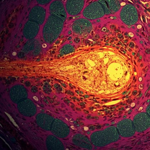
Single-cell analysis of two severe COVID-19 patients reveals a monocyte-associated and tocilizumab-responding cytokine storm
C. Guo, H. Ma, et al.
This cutting-edge study by Chuang Guo, Huan Ma, and colleagues delves into single-cell analysis of severe COVID-19 patients, revealing a specific monocyte subpopulation linked to cytokine storms. The research showcases how tocilizumab effectively mitigates inflammation while preserving vital immune responses, paving the way for better treatment strategies against COVID-19's severe complications.
Related Publications
Explore these studies to deepen your understanding
Adjacent work that informs or extends this paper's methodology and findings.

Medicine and Health
Single-cell analysis of chromatin and expression reveals age- and sex-associated alterations in the human heart
D. F. Read, G. T. Booth, et al.

Medicine and Health
Effects of exercise therapy on anxiety and depression in patients with COVID-19: a systematic review and meta-analysis
J. Tang, L. Chen, et al.

Medicine and Health
Critical roles of cytokine storm and bacterial infection in patients with COVID-19: therapeutic potential of mesenchymal stem cells
B. Arjmand, S. Alavi-moghadam, et al.

Medicine and Health
Prevalence and prognosis of tinnitus in post-COVID-19 patients: a cross-sectional survey
S. Mao, D. Gu, et al.


