
Medicine and HealthMucosal Immunology
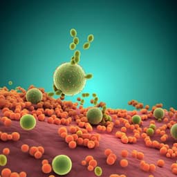
Perturbation of the gut microbiome by *Prevotella* spp. enhances host susceptibility to mucosal inflammation
A. Iljazovic, U. Roy, et al.
This groundbreaking study reveals how colonization of mice with Prevotella intestinalis worsens intestinal inflammation by modifying the gut microbiome and reducing vital short-chain fatty acids. Notably, IL-18 levels drop significantly, leading to increased inflammation. However, supplementation with IL-18 shows promise in mitigating this response. These insights from Aida Iljazovic and colleagues could reshape our understanding of metabolic shifts in gut health.
Related Publications
Explore these studies to deepen your understanding
Adjacent work that informs or extends this paper's methodology and findings.
Medicine and Health
Targeted delivery of the probiotic *Saccharomyces boulardii* to the extracellular matrix enhances gut residence time and recovery in murine colitis
M. K. Heavey, A. Hazelton, et al.

Medicine and Health
Impact of the gut microbiome on immunological responses to COVID-19 vaccination in healthy controls and people living with HIV
S. Ray, A. Narayanan, et al.

Business
Not with the bot! The relevance of trust to explain the acceptance of chatbots by insurance customers
J. D. Andrés-sánchez and J. Gené-albesa
Medicine and Health
Histones released by NETosis enhance the infectivity of SARS-CoV-2 by bridging the spike protein subunit 2 and sialic acid on host cells
W. Hong, J. Yang, et al.


