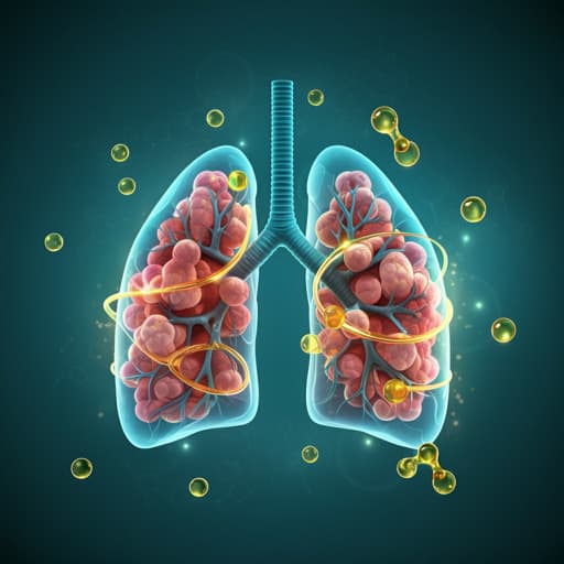
Omega-3 polyunsaturated fatty acids ameliorate PM2.5 exposure induced lung injury in mice through remodeling the gut microbiota and modulating the lung metabolism
J. Li, Y. Chen, et al.
Explore these studies to deepen your understanding
Adjacent work that informs or extends this paper's methodology and findings.

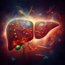
The interplay between dietary fatty acids and gut microbiota influences host metabolism and hepatic steatosis
M. Schoeler, S. Ellero-simatos, et al.

The effects of omega 3 fatty acids on the serum concentrations of pro inflammatory cytokines and depression status in patients with bipolar disorder: A randomized double-blind controlled clinical trial
H. Eslahi, M. Shakiba, et al.
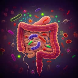
Ginger essential oil prevents NASH progression by blocking the NLRP3 inflammasome and remodeling the gut microbiota-LPS-TLR4 pathway in mice
S. Panyod, W. Wu, et al.
Ablation of the gut microbiota alleviates high-methionine diet-induced hyperhomocysteinemia and glucose intolerance in mice
W. Li, Y. Jia, et al.


