
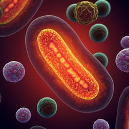
Mitochondrial Oxidative Stress Is the General Reason for Apoptosis Induced by Different-Valence Heavy Metals in Cells and Mitochondria
S. M. Korotkov
Explore these studies to deepen your understanding
Adjacent work that informs or extends this paper's methodology and findings.
N-acetylcysteine supplementation did not reverse mitochondrial oxidative stress, apoptosis, and inflammation in the salivary glands of hyperglycemic rats
Z. Anna, K. Joanna, et al.
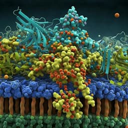
MFRP is a molecular hub that organizes the apical membrane of RPE cells by engaging in interactions with specific proteins and lipids
A. Tworak, R. Smidak, et al.

Dopamine release and dopamine-related gene expression in the amygdala are modulated by the gastrin-releasing peptide in opposite directions during stress-enhanced fear learning and extinction
Y. Morishita, I. Fuentes, et al.

Dopamine release and dopamine-related gene expression in the amygdala are modulated by the gastrin-releasing peptide in opposite directions during stress-enhanced fear learning and extinction
Y. Morishita, I. Fuentes, et al.


