Medicine and Health
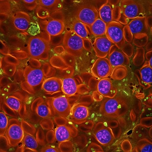
Menstrual blood-derived stromal cells modulate functional properties of mouse and human macrophages
R. Martínez-aguilar, S. Romero-pinedo, et al.
This study reveals the intriguing effects of menstrual blood-derived stromal cells (MenSCs) on macrophage populations during acute inflammation. Conducted by Rocío Martínez-Aguilar and colleagues, the research shows how MenSCs can modulate immune responses, potentially influencing future clinical applications.
~3 min • Beginner • English
Introduction
Menstrual blood-derived stromal cells (MenSCs) are mesenchymal stromal cells (MSCs) isolated from menstrual fluid that share core MSC properties, including adherence, expression of mesenchymal markers, trilineage differentiation in vitro, and low immunogenicity. Their non-invasive collection and abundance position MenSCs as promising candidates for clinical cell-based therapies. While MSCs are recognized for paracrine-mediated immunomodulatory actions, the specific effects of MenSCs on innate immune populations, particularly macrophages that orchestrate inflammation onset and maintenance, are not fully defined. This study aims to determine how MenSCs influence macrophage recruitment and function during early acute inflammation using two mouse peritonitis models (sterile TGC-elicited and Salmonella Typhimurium-induced sepsis), and to examine direct effects on human macrophage-like cells in vitro, focusing on phagocytosis, bacterial clearance, and reactive oxygen species production. Understanding these mechanisms is critical for optimizing MenSC-based therapies for inflammatory diseases.
Literature Review
Extensive literature describes MSC immunomodulatory activity via secreted cytokines, anti-inflammatory factors, and cell-cell contact. Tissue source can influence MSC functional properties, necessitating caution when extrapolating effects across MSC types. Prior MenSC studies report benefits in experimental colitis, LPS-induced injury, and polymicrobial sepsis, often emphasizing systemic outcomes (e.g., survival, histopathology) or adaptive immune effects rather than direct innate cell modulation. MSCs from other sources can polarize macrophages toward regulatory phenotypes and enhance phagocytosis, yet the impact of MenSCs specifically on macrophage recruitment and bactericidal function remains unclear. This gap motivates the present work to dissect MenSC-macrophage crosstalk in controlled inflammatory contexts.
Methodology
Ethics: Human sample collection and animal studies were approved by University of Granada committees (human: 186/CEIH/2016; animal: 12/12/2016-179/143-CEEA-OH-2015 and 12/12/2016-179 JJAA). Human MenSC isolation and culture: Menstrual blood from 15 healthy women (20–35 years) was collected on cycle day 2 using cups/tampons. Mononuclear cells were isolated by Ficoll-Paque density centrifugation and cultured in Opti-MEM with antibiotics and 3% FBS at 37 °C, 5% CO2. Early passages (P2–P6) were used. MenSC characterization included morphology, trilineage differentiation (not shown), and flow cytometry phenotype: positive for CD9, CD29, CD44, CD73, CD90, CD105, CD146; low CD117; negative for CD14, CD34, CD38, CD45, CD133, HLA-DR. THP-1 culture and differentiation: THP-1 cells were grown in supplemented RPMI 1640; differentiated to macrophage-like cells with 10 ng/ml PMA for 24 h, then rested 24 h in Opti-MEM (3% FBS). For co-cultures, THP-1PMA (4×10^5/well) and MenSCs (1×10^5/well; 4:1 ratio) were cultured in Transwells (0.4 µm) for 48–72 h depending on assay. Bacterial strains: Salmonella enterica serovar Typhimurium wild-type (NCTC 12023) and attenuated SseB mutant (MvP643, p3232; ampicillin resistant) were grown in LB (±100 µg/ml ampicillin). Animals: Female BALB/c mice (8–10 weeks) were housed under standard conditions. TGC-elicited peritonitis model: Mice received 4% thioglycollate intraperitoneally (IP). MenSCs (2×10^6) were injected IP at 4 h or 24 h post-TGC. Controls: PBS, TGC alone, MenSCs alone. Mice were euthanized 4 days after TGC; peritoneal lavages were performed (3×8 ml cold PBS), cells counted and phenotyped by flow cytometry (macrophages F4/80+CD11b+, neutrophils Ly-6G+CD11b+, inflammatory monocytes Ly-6C+CD11b+). Monobacterial sepsis model: Mice were IP challenged with 2.5×10^6 wild-type Salmonella Typhimurium (ST) + 2.5×10^6 SseB. MenSCs (2×10^6) were injected IP 4 h later. Mice were euthanized 24 h after infection. Outcomes: peritoneal cell counts and phenotypes; body weight change; spleen weight relative to body weight; bacterial loads (CFU) in spleen, mesenteric lymph nodes, and peritoneal fluid plated on LB and LB+ampicillin; peritoneal fluid mRNA expression (Nos2, Tnf, Il10, Tgfb) by qPCR normalized to Actb using 2^-ΔΔCT. Histology/immunofluorescence: Peritoneal aggregates (from TGC+MenSCs-24 h) were processed for H&E (paraffin) and confocal microscopy (OCT, cryosections). Staining included anti-human nuclei (MenSCs), anti-F4/80 (macrophages), and anti-Ly-6G (neutrophils). Flow cytometry: Cells stained with specific antibodies (Fc-block used where appropriate); data acquired on FACSCalibur or FACSCanto II and analyzed with FlowJo/FACSDiva. In vitro functional assays: - Phagocytosis assay: After 48 h THP-1PMA–MenSCs Transwell co-culture, MenSC inserts removed; THP-1PMA infected with Salmonella SseB at MOI 25 for 30 or 60 min; extracellular bacteria washed; cells lysed; serial dilutions plated on LB+ampicillin to count intracellular CFU. - Gentamicin protection (killing) assay: Following 48 h co-culture, THP-1PMA infected with Salmonella SseB (MOI 25) for 1 h; treated with gentamicin 100 µg/ml for 1 h then 10 µg/ml; cells lysed at 6, 24, 48 h; CFU enumerated. PFA-fixed cells served as killing-negative controls. - Intracellular ROS: THP-1PMA cultured ± direct MenSC co-culture (4:1) for 48 h; loaded with DCFDA (3 µM) 1 h; stimulated with Salmonella SseB (MOI 100); fluorescence measured over time. Statistics: Mean±SEM; TGC model analyzed by one- or two-way ANOVA with Tukey’s post hoc; sepsis model by unpaired two-tailed t-test (weights, total cells) and two-way ANOVA with Sidak’s post hoc (phenotypes, CFU, mRNA); in vitro by two-way ANOVA with Tukey’s/Sidak’s as appropriate. Significance p<0.05.
Key Findings
- In TGC-elicited peritonitis, MenSC timing influenced recruitment: • Total peritoneal cells were unchanged when MenSCs were given at 4 h post-TGC (T-MenSC-4 h: 1.1×10^7±4.2×10^5) vs TGC alone (1.1×10^7±5.6×10^5), but significantly reduced when given at 24 h (T-MenSC-24 h: 3.5×10^6±4.7×10^5; p<0.0001). • Macrophage numbers (F4/80+CD11b+) decreased vs TGC (7.4×10^6±5×10^5): T-MenSC-4 h 6.1×10^6±8.2×10^5 (p<0.01); T-MenSC-24 h 8.2×10^5±1.5×10^5 (p<0.0001). • Neutrophil numbers (Ly-6G+CD11b+) increased in T-MenSC-4 h (3.1×10^6±1.5×10^5; p<0.0001) vs TGC (8×10^5±1.7×10^5); T-MenSC-24 h similar to TGC (4×10^5±1×10^5 vs 8×10^5±1.7×10^5). • Inflammatory monocyte numbers (Ly-6C+CD11b+) showed no significant changes among groups. • Proportions shifted: macrophages decreased (TGC 84%±2.3; T-MenSC-4 h 58%±4.4, p<0.0001; T-MenSC-24 h 37%±3.3, p<0.0001); neutrophils increased (TGC 6%±1.6; 4 h 26%±1.8, p<0.0001; 24 h 18%±2.2, p<0.01); inflammatory monocytes increased at 24 h (11%±1.9 vs 2%±0.8; p<0.05). MenSCs alone reduced macrophage percentage vs PBS (46%±5.7 vs 80%±4.5; p<0.0001). • Peritoneal nodular aggregates formed in T-MenSC-24 h mice. Histology showed organized structures with human MenSCs (anti-human nuclei+) within the aggregates, macrophages (F4/80+) predominantly peripheral and perivascular, and neutrophils (Ly-6G+) present within the structures. - In Salmonella-induced sepsis, MenSCs reduced peritoneal phagocyte recruitment and worsened bacterial control: • Total peritoneal cell counts and both macrophage (F4/80+CD11b+) and neutrophil (Ly-6G+CD11b+) numbers and percentages were decreased 24 h after MenSC administration compared to infection alone. • Mice receiving MenSCs exhibited greater bacterial burdens (CFU) in spleen, peritoneal fluid, and mesenteric lymph nodes for both wild-type ST and attenuated SseB strains, indicating impaired bacterial clearance and increased dissemination. • Peritoneal fluid mRNA showed increased Nos2 and Il10 expression in MenSC-treated mice relative to infected controls, while Tnf and Tgfb showed no significant upregulation, suggesting a shift toward a regulatory macrophage phenotype. • Body weight and spleen weight metrics were consistent with worsened infection in MenSC-treated animals (directional changes reported; statistics applied as specified). - In vitro co-culture with human macrophage-like THP-1PMA cells revealed direct functional reprogramming by MenSCs: • Phenotype: Increased CD14 expression (shift from CD14low to CD14high) and reduced co-stimulatory molecules CD80/CD86 on THP-1PMA after 72 h MenSC co-culture, consistent with enhanced phagocytic but more regulatory/less antigen-presenting phenotype. • Phagocytosis: Uptake of Salmonella SseB increased at 30 and 60 min in MenSC-exposed THP-1PMA vs controls. • Bacterial killing: Gentamicin protection assays showed higher intracellular CFU at 6, 24, and 48 h in MenSC-exposed THP-1PMA, indicating impaired killing. • ROS: DCFDA measurements revealed significantly reduced ROS production across time in THP-1PMA co-cultured directly with MenSCs, correlating with diminished bactericidal activity, despite MenSCs themselves being capable of generating ROS upon bacterial challenge. Overall, MenSCs decreased macrophage recruitment and bactericidal function and increased phagocytosis while reducing ROS, leading to poorer bacterial clearance in vivo.
Discussion
This work demonstrates that MenSCs modulate macrophage-mediated innate responses in a context-dependent manner. In sterile TGC peritonitis, MenSCs reduced macrophage recruitment, increased neutrophil proportions, and induced peritoneal aggregates where MenSCs localized centrally and macrophages peripherally, suggesting MenSCs act as hubs that attract and organize innate cells, potentially via chemokines such as CCL2. In infectious peritonitis, MenSC administration reduced recruitment of macrophages and neutrophils and coincided with increased bacterial loads and dissemination, indicating impaired compartmentalization and clearance of bacteria. Elevated Nos2 and Il10 in peritoneal fluid supports a shift toward regulatory macrophage features, which are associated with reduced bactericidal capacity and limited neutrophil infiltration. In vitro, MenSCs increased macrophage phagocytic receptor expression (CD14) and phagocytosis but reduced co-stimulatory markers (CD80/CD86), ROS generation, and bacterial killing, reinforcing a regulatory reprogramming of macrophages. Collectively, MenSCs appear to act as sensors and switchers of inflammation, dampening damaging inflammation and promoting homeostasis; however, in bacterial infection such modulation may compromise microbial clearance. These findings refine the understanding of MenSC mechanisms with implications for their clinical use in inflammatory disorders, where timing, inflammatory context (DAMP- vs PAMP-driven), and infection status are likely critical determinants of therapeutic outcomes.
Conclusion
The study establishes that MenSCs significantly modulate macrophage recruitment and function in acute inflammation. In sterile conditions, MenSCs reduce macrophage influx and organize peritoneal immune aggregates, while in bacterial sepsis they diminish phagocyte recruitment and impair bacterial clearance, likely via induction of a regulatory macrophage phenotype characterized by increased CD14 and IL-10, reduced CD80/CD86, and diminished ROS. In vitro, MenSCs enhance phagocytosis but reduce bactericidal activity of human macrophage-like cells. These context-dependent effects should be carefully considered for clinical translation of MenSC-based therapies. Future research should identify the soluble mediators and signaling pathways mediating MenSC-macrophage crosstalk, define how timing and inflammatory cues shape MenSC responses, and extend analyses to other innate cells (e.g., neutrophils) and to primary human macrophages to optimize therapeutic strategies.
Limitations
The study does not identify the specific mediators or molecular pathways by which MenSCs reprogram macrophages. Innate cell analyses focused primarily on macrophages, with limited mechanistic assessment of neutrophils and other populations. In vivo findings are model- and strain-specific (BALB/c; Salmonella Typhimurium co-infection with attenuated SseB), which may limit generalizability to other sepsis models or pathogens. In vitro macrophage data rely on PMA-differentiated THP-1 cells rather than primary human macrophages. The observed peritoneal aggregates were characterized in limited numbers, and causal chemokines/cell trafficking mechanisms were not directly tested.
Related Publications
Explore these studies to deepen your understanding of the subject.

