Medicine and Healthnpj Regenerative Medicine
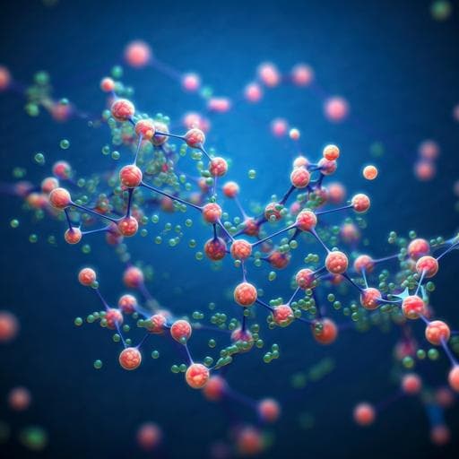
Injectable decellularized cartilage matrix hydrogel encapsulating urine-derived stem cells for immunomodulatory and cartilage defect regeneration
J. Zeng, L. Huang, et al.
This groundbreaking study unveils an innovative injectable hydrogel derived from pig cartilage, aimed at repairing cartilage defects. The research, conducted by an expert team, including Junfeng Zeng and Liping Huang among others, demonstrates the hydrogel's impressive ability to enhance the chondrogenic differentiation of human urine-derived stem cells both in lab conditions and in a rat model, paving the way for advanced cartilage regeneration strategies.
Related Publications
Explore these studies to deepen your understanding
Adjacent work that informs or extends this paper's methodology and findings.
Medicine and Health
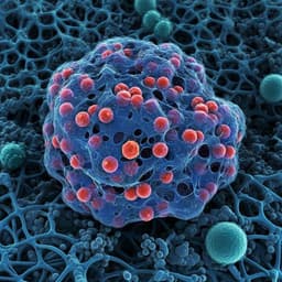
Highly porous and injectable hydrogels derived from cartilage acellularized matrix exhibit reduction and NIR light dual-responsive drug release properties for application in antitumor therapy
M. Gulfam, S. Jo, et al.
Medicine and Health
An injectable photopolymerized hydrogel with antimicrobial and biocompatible properties for infected skin regeneration
A. Sun, X. He, et al.
Medicine and Health
Ultra-durable cell-free bioactive hydrogel with fast shape memory and on-demand drug release for cartilage regeneration
Y. Yang, X. Zhao, et al.
Medicine and Health
An instantly fixable and self-adaptive scaffold for skull regeneration by autologous stem cell recruitment and angiogenesis
G. Lu, Y. Xu, et al.


