
Medicine and Health
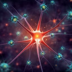
Brain-derived neurotrophic factor from microglia regulates neuronal development in the medial prefrontal cortex and its associated social behavior
T. Komori, K. Okamura, et al.
This groundbreaking study by Takashi Komori and colleagues reveals how microglia-derived BDNF impacts social behavior and mPFC development in mice. Explore the complex relationship between childhood experiences and neurobiology, uncovering vital insights into sociability and brain function.
~3 min • Beginner • English
Introduction
Microglia refine synapses in an activity-dependent manner and are essential for normal brain development and critical-period plasticity. Similar to sensory functions, social behaviors in mice develop via experience-dependent maturation during a limited postnatal window, with the medial prefrontal cortex (mPFC) being a key region regulating social behaviors. Juvenile social deprivation disrupts neural circuits and glial functions in the mPFC and results in lasting social deficits. Prior work implicates microglia in social development, synaptic pruning in the prefrontal cortex, and time-specific maturation of the mPFC. Brain-derived neurotrophic factor (BDNF) maintains inhibitory interneurons and contributes to closing critical periods. Microglia secrete BDNF, which can modulate synaptic plasticity. However, whether microglia-secreted BDNF (MG-BDNF) influences cortical critical-period plasticity and social behavior development was unknown. This study tests the hypothesis that MG-BDNF regulates time-specific mPFC circuit development and adult social behaviors, and examines whether modulating MG-BDNF during the juvenile critical period versus later periods has distinct outcomes.
Literature Review
- Microglia shape developing brain circuits and prune synapses in an activity-dependent manner; their role has been linked to critical period plasticity.
- Social behavior development is experience dependent and requires juvenile social experience for normal adult sociability; juvenile social deprivation disrupts mPFC circuits and glial cell function and causes persistent social impairments.
- Microglia contribute to time-specific pruning and functional maturation of the prefrontal cortex, including the mPFC.
- BDNF supports inhibitory interneuron maintenance and critical period closure; microglia are a source of BDNF. BDNF has been implicated in learning-dependent synaptic plasticity in cortex and inhibitory transmission in spinal cord.
- Prior to this work, the role of microglia-derived BDNF in cortical critical-period plasticity and social behavior development was unclear, motivating the current time-specific manipulations of MG-BDNF.
Methodology
Animals and housing: Male mice (C57BL/6J background with mixed 129/Sv in transgenic lines) were housed under standard conditions (12-h light/dark, food and water ad libitum). Juvenile social isolation (j-SI) was induced by single housing from P21 to P35, followed by regrouping; controls were group-housed.
Transgenic models: Iba1-tTA transgenic mice were crossed with tetracycline operator Bdnf knock-in mice to generate microglia-specific Bdnf-overexpressing mice (Iba1-tTA(+/-):Bdnf(tetO/tetO)) and Bdnf(tetO/tetO) control mice. Temporal control of BDNF overexpression was achieved by doxycycline (DOX)-containing chow (100 mg/kg) administered beginning at specified ages (e.g., from P21 to suppress during juvenile period; or initiated at P45–P50 for delayed normalization).
Behavioral assays: Three-chamber social preference test to assess sociability; open field test for locomotion/anxiety-like behavior; augmented reality-based long-term animal behavior observation system (AR-LAB) to quantify naturalistic social approaches among multiple unfamiliar mice.
Molecular assays: RT-qPCR for Bdnf mRNA in microglia isolated from cortex and mPFC; ELISA for BDNF protein in microglia; bulk tissue RT-qPCR; RNA-sequencing (mPFC) with PCA, differential expression analysis, hierarchical clustering, and Gene Ontology analyses; assessment of complement system-related genes (e.g., C1qa, C1qb, C1qc, C3ar1) with Z-scores.
Electrophysiology: Whole-cell patch-clamp recordings from mPFC layer V pyramidal neurons to measure excitability (spike frequency, amplitude, threshold), spontaneous EPSCs/IPSCs (sEPSCs/sIPSCs), miniature EPSCs/IPSCs (mEPSCs/mIPSCs). Additional recordings in dorsolateral PFC to assess regional specificity.
Statistics: Normality testing (Shapiro–Wilk), variance (Bartlett’s), unpaired two-tailed t-tests or Mann–Whitney U tests as appropriate; Kruskal–Wallis for validation; two-way ANOVA (e.g., time x phenotype), two-way repeated measures ANOVA for cumulative behaviors; Bonferroni post hoc tests; Spearman correlations for expression-behavior/imaging and human CATS-BDNF analyses; Benjamini–Hochberg FDR control. Significance set at p<0.05 (q where applicable).
Key Findings
- Juvenile social isolation (P21–P35) reduced adult sociability without affecting open field activity and increased Bdnf mRNA expression specifically in microglia from cortex and mPFC.
- Three-chamber sociability: j-SI mice had lower social interaction score than GH controls (t(20)=3.94, p=0.0005) and spent less time near a novel mouse (F(2,18)=7.89, p=0.003; Bonferroni post hoc GH vs j-SI p<0.0001).
- Microglial Bdnf RT-qPCR: cortex higher in j-SI vs GH (t(22)=4.876, p<0.0001); mPFC trend reported.
- Sustained microglial Bdnf overexpression (Iba1-tTA(+/-):Bdnf(tetO/tetO)) recapitulated social deficits and altered mPFC physiology.
- Microglial BDNF protein increased by ELISA.
- Three-chamber: lower social interaction score vs control (Mann–Whitney U, t=4.7, p=0.0226); less time near novel mice (two-way ANOVA F(3,13)=5.9, p=0.0001; Bonferroni significant). No open field differences.
- AR-LAB: fewer approaches to other mice across time (two-way ANOVA: interaction F(3,21)=3.926, p<0.0001; phenotype effect F(1,21)=6.676, p=0.0158; time effect F(3,21)=39.31, p<0.0001); total activity unchanged (p=0.4276).
- Electrophysiology in mPFC layer V pyramidal neurons indicated increased inhibitory inputs and altered excitability with MG-BDNF overexpression.
- sEPSC frequency increased (*) with unchanged amplitude; sIPSC frequency increased (***) with unchanged amplitude.
- mEPSC frequency increased (***) with unchanged amplitude; mIPSC frequency (***) and amplitude (*) increased.
- Spike properties: reduced spike frequency (****), altered amplitude (**), spike width not significantly changed (ns).
- No significant changes detected in dorsolateral PFC, supporting regional specificity to mPFC.
- Transcriptomics implicated complement and Wnt-related functions.
- In adult MG-BDNF-overexpressing mPFC: 63 genes downregulated and 39 upregulated vs controls; GO molecular functions enriched for Wnt-activated receptor activity (corrected p<0.0001), Wnt-protein binding (corrected p<0.001), and others.
- Complement system-related genes showed reduced Z-scores; significantly decreased C1qa (corrected p=0.0241) and C3ar1 (corrected p=0.0288), suggesting reduced complement cascade activity.
- Time-specific normalization of MG-BDNF with doxycycline differentially rescued outcomes.
- Normalization from P21 (juvenile period) restored sociability and normalized mPFC electrophysiological measures (spike frequency/amplitude and s/m EPSC/IPSC frequencies comparable to controls).
- Normalization initiated at P45–P50 improved sociability and restored some excitatory measures (e.g., eEPSC/mEPSC frequencies) but failed to normalize reduced spike frequency and increased sIPSC/mIPSC frequencies; complement system-related gene expression changes were restored.
- Human translational correlate: In peripheral monocyte-derived macrophages, BDNF expression correlated with adverse childhood experiences.
- No correlation in M1 macrophages (rs=0.0694, p=0.6746, N=39).
- Significant positive correlation in M2 macrophages between total CATS score and BDNF expression (rs=0.4088, p=0.00098, N=39); FDR-adjusted q=0.033 indicated significance for total and certain subscales.
Discussion
The study demonstrates that microglia-derived BDNF (MG-BDNF) critically shapes mPFC circuit development linked to social behaviors. Juvenile social isolation elevates microglial Bdnf and leads to enduring sociability deficits. Experimentally elevating MG-BDNF was sufficient to reproduce social impairments and to bias mPFC layer V pyramidal neurons toward increased inhibitory synaptic input and reduced excitability. These physiological alterations map onto a disrupted developmental trajectory of the mPFC.
Temporal manipulations revealed a critical period: normalizing MG-BDNF beginning at P21 rescued both sociability and multiple electrophysiological parameters, whereas normalization beginning at P45–P50 restored sociability and some excitatory measures but left inhibitory synaptic abnormalities and reduced spike frequency. This dissociation suggests that adult social behavior deficits induced by MG-BDNF rely primarily on reduced excitatory tone in mPFC, while inhibitory synaptic excess may be less directly coupled to sociability or may require earlier intervention to normalize.
Transcriptomic changes point to MG-BDNF-mediated modulation of complement and Wnt-related pathways. Decreases in C1qa and C3ar1 suggest reduced microglial complement signaling, potentially diminishing activity-dependent synaptic pruning of both excitatory and inhibitory synapses and contributing to the excitatory/inhibitory imbalance. Restoration of complement gene expression by adult normalization (P45–P50) without full electrophysiological rescue implies complement-mediated maturation is predominantly juvenile.
The human macrophage findings provide a translational link, indicating that adverse childhood experiences correlate with elevated BDNF in M2 macrophages, cells sharing functional features with microglia. Together, these findings position MG-BDNF as a time-sensitive regulator of mPFC circuitry underlying social behavior, with potential relevance to how early-life experiences shape adult socio-affective outcomes.
Conclusion
Microglia-derived BDNF is a key, time-sensitive regulator of mPFC circuit maturation and adult social behavior. Juvenile social isolation elevates microglial Bdnf and impairs sociability; sustained experimental MG-BDNF overexpression recapitulates social deficits and induces increased inhibitory input and reduced excitability in mPFC pyramidal neurons. Normalizing MG-BDNF from the juvenile period rescues both behavioral and electrophysiological phenotypes, whereas later normalization rescues sociability but not all circuit abnormalities. MG-BDNF overexpression alters complement-related gene expression, implicating microglial complement signaling in developmental synaptic refinement. In humans, BDNF expression in M2 macrophages correlates positively with adverse childhood experiences, suggesting a translational link between early-life experience and immune-derived neurotrophic signaling.
Future directions include: dissecting cell-type-specific contributions (microglia vs astrocytes) to BDNF signaling; mapping regional specificity beyond mPFC; mechanistically linking MG-BDNF to complement-mediated synaptic pruning; and longitudinal human studies to clarify peripheral macrophage BDNF as a biomarker of early-life adversity and mPFC-related social function.
Limitations
- Genetic background: Control and experimental mice derive from Bdnf(tetO) lines with mixed C57BL/6J and 129/Sv backgrounds; although balanced across groups, residual background effects cannot be fully excluded.
- Timing vs duration: While timing of DOX initiation (P21 vs P45–P50) showed distinct outcomes, the total duration of MG-BDNF suppression may also influence results.
- Cell-type specificity: Astrocytes also regulate BDNF signaling and could contribute to behavioral phenotypes; MG-BDNF effects cannot exclude astrocytic involvement.
- Regional scope: Electrophysiological analyses focused on mPFC (and a check in dorsolateral PFC); broader brain-region assessments are needed.
- Complement attribution: Complement gene changes were measured in bulk mPFC; definitive attribution to microglia vs neurons/glia is limited.
- Mouse-human translation: Peripheral macrophage BDNF may not reflect brain microglial BDNF; correlations with human childhood experiences are associative and not causal.
Related Publications
Explore these studies to deepen your understanding of the subject.