logo
ResearchBunny Logo
A diet high in sugar and fat influences neurotransmitter metabolism and then affects brain function by altering the gut microbiota

Medicine and Health

A diet high in sugar and fat influences neurotransmitter metabolism and then affects brain function by altering the gut microbiota

Y. Guo, X. Zhu, et al.

Discover how gut microbiota metabolites influence brain function and neurotransmitter metabolism in a groundbreaking study by Yinrui Guo and colleagues. Using a high-sugar, high-fat diet to induce gut dysbiosis in mice, the research uncovers a novel connection between metabolism, brain circRNAs, and gut health, paving the way for new therapeutic insights.

00:00
00:00
~3 min • Beginner • English
Introduction
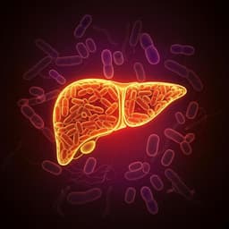
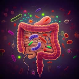
The study investigates how a high-sugar, high-fat (HSHF) diet perturbs the gut microbiota (GM) and how this disturbance affects neurotransmitter metabolism and brain function via the gut–brain axis. GM metabolites influence immunity and metabolism and can act locally in the gut or reach systemic organs, including the brain. Prior work implicates the GM in neurodegenerative diseases such as Alzheimer’s and Parkinson’s, and suggests that microbiota-derived neuroactive molecules can regulate host behavior and physiology. Diet-induced dysbiosis (e.g., from high fat/sugar intake and low fiber) increases intestinal permeability and alters dominant bacterial populations, potentially enabling bacteria to produce or modulate neurotransmitters that affect host neural circuits. The authors aim to: (1) perturb GM using an HSHF diet, (2) measure ensuing changes in pathology, neurotransmitters, metabolism, and brain circular RNA (circRNA) transcription, and (3) identify links among GM, neurotransmitter metabolism, and brain function, expanding a “microbiome–transcriptome” linkage library relevant to gut–brain communication and therapeutic development.
Literature Review
Methodology
Animal models and diets: Adult male KM mice (18–22 g, 6 weeks) were randomized to control (standard chow) or HSHF diet groups (n=12/group) for 3 months, housed at 25±2 °C, 12-h light–dark cycle, food/water ad libitum. HSHF diet composition: 20% sucrose, 15% fat, 12% cholesterol, 0.2% bile acid sodium, 10% casein, 0.6% calcium hydrogen phosphate, 0.4% stone powder, 0.4% premix, 52.5% basal feed; energy ratio: protein 17%, fat 17%, carbohydrate 46%. TMA exposure in rats: Male Sprague–Dawley rats (180–220 g) were assigned to normal or trimethylamine (TMA)-treated groups (n=10/group). TMA group received 2 mL/kg of 2.5% TMA. Housing conditions matched mice. Pathogen administration to probe neurotransmitter modulation: Adult male C57 mice (18–22 g) were divided into four groups (n=5/group): normal, Candida albicans-treated, Klebsiella pneumoniae-treated, and combined C. albicans + K. pneumoniae (Ca+Kp). Pathogens were administered intragastrically as monotherapy or in combination to assess effects on the choleric/cholinergic system and neurotransmitter metabolism. Physiological and biochemical measurements: Daily monitoring of appearance, behavior, and intake; bodyweight measured every 3 days. At endpoint, blood collected for glucose (BG), triglycerides (TG), total cholesterol (T-CHOL), and HDL-C using commercial kits. Serum and brain tissue levels of trimethylamine-N-oxide (TMAO) and multiple neurotransmitters quantified by LC-MS. Histopathology and immunostaining: Brain, liver, kidney, spinal cord, spleen, and adipose tissues fixed in 4% paraformaldehyde, processed into paraffin sections. Brain sections stained with H&E, silver, Nissl, and TUNEL; immunostaining performed using a peroxidase-conjugated polymer kit (DAKO Envision). Light microscopy used for assessment of obesity-related and other pathological changes. Microbiome profiling: Intestinal content collected and stored at −80 °C. Microbial DNA extracted (1.2–20.0 ng total), 16S rRNA genes amplified using primers 5'-ACTCCTACGGGAGGCAGCAG-3' (forward) and 5'-GACGTACCHVGGGTWTCTAAT-3' (reverse). PCR products purified, quantified, pooled, and sequenced on Illumina HiSeq 2500. Bioinformatic processing performed to profile GM composition. Targeted metabolomics of neurotransmitters: Standards prepared and benzoyl chloride (BZ) derivatization used; internal standard with 13C2-labeled BZ. Calibration range covered 0.001–1200 ng/mL. Brain tissue (20±1 mg) homogenized in ascorbic acid solution, extracted with prechilled acetonitrile, derivatized with BZ, and analyzed by UPLC–MS/MS; dilution strategies applied for high vs. low concentration analytes. Intestinal content processed similarly with ascorbic acid and acetone extraction, BZ derivatization, and UPLC–MS/MS. UPLC–MS/MS conditions: Waters Acquity UPLC with BEH C18 column (100×2.1 mm, 1.7 µm), 0.4 mL/min, mobile phase A: 2 mM ammonium acetate with 0.1% formic acid in water; B: acetonitrile; gradient from 10% to 90% B and re-equilibration over 10 min; column at 40 °C. AB Sciex Triple Quad 5500 in positive ESI: 600 °C source, curtain gas 30 psi, gas1 50 psi, gas2 50 psi, collision gas 8 psi, 5500 V, entrance potential 10 V, CCE 14 V. Scheduled MRM used; data acquired with Analyst v1.5.2; peak integration for quantification. RNA isolation and sequencing: Total RNA isolated (QIAzol + RNeasy), DNase-treated; rRNA depleted (Ribo-Zero), linear RNA digested with RNase R to enrich circRNAs. cDNA libraries prepared (Illumina TruSeq RNA Sample Preparation), sequenced on Illumina HiSeq 2500. Computational analysis: Reads mapped to UCSC transcript set with Bowtie2 v2.1.0; expression estimated by RSEM v1.2.15; lincRNA expression from Lincpedia; TMM normalization; differentially expressed genes defined by edgeR (p<0.05 and >1.5-fold change); pathway analysis using KEGG with Fisher’s exact test and FDR correction. For circRNAs, reads mapped with STAR; circRNA identification/quantification with DCC; TMM normalization; DE analysis with edgeR; miRNA targets predicted with miRanda; visualization in R. RT-qPCR validation: circRNA candidates validated by RT-qPCR using outward-facing primers specific to circRNA back-splice junctions and inward primers for linear transcripts (primer design per Memczak; sequences in Table 2).
Key Findings
- An HSHF diet induced gut dysbiosis, damaged the intestinal tract, altered neurotransmitter metabolism in both intestine and brain, and led to changes in brain function and brain circRNA profiles. - The gut microbial metabolite trimethylamine-N-oxide (TMAO) was capable of degrading some brain circRNAs, linking microbial metabolism to transcriptomic regulation in the brain. - The baseline composition of the gut microbiota determined the conversion rate of choline to TMAO, indicating host–microbiome variability in metabolite production. - Altering the abundance of a single bacterial strain could influence neurotransmitter secretion, supporting a causal role for specific microbes in modulating host neurochemistry. - Together, results suggest a mechanistic connection among GM composition, metabolite (TMA/TMAO) dynamics, neurotransmitter homeostasis, and brain transcriptomic changes (circRNAs).
Discussion
The study demonstrates that dietary perturbation of the gut microbiota via an HSHF diet can propagate changes along the gut–brain axis, manifesting as disrupted intestinal integrity, altered neurotransmitter metabolism locally and centrally, and shifts in brain circRNA expression. The identification of TMAO as a factor that can degrade certain circRNAs provides a plausible biochemical route by which microbial metabolites affect brain transcriptomic regulation. The finding that baseline GM composition modulates choline-to-TMAO conversion explains inter-individual variability in metabolite exposure and its downstream neural consequences. Moreover, showing that changes in the abundance of a single microbial strain can alter neurotransmitter secretion supports a causal microbial influence on host neurochemistry. Collectively, these observations address the central hypothesis that GM alterations can influence brain function via metabolic and transcriptomic pathways, and highlight potential therapeutic levers—diet, microbial composition, and metabolite modulation—for neuro-related conditions.
Conclusion
This work links diet-induced gut dysbiosis to altered neurotransmitter metabolism and brain function, with concomitant changes in brain circRNA profiles. It identifies TMAO as a microbial metabolite capable of degrading specific circRNAs and underscores that baseline GM composition governs choline-to-TMAO conversion. The results expand the “microbiome–transcriptome” linkage library and offer insights into gut–brain communication with potential implications for biomarkers and therapeutic strategies in pathology, toxicology, diet, and nutrition. Future studies should define the specific circRNAs and neural pathways affected, establish causal relationships with behavior and neurodegeneration models, and evaluate targeted microbiota or dietary interventions to modulate metabolite and neurotransmitter profiles.
Limitations
Listen, Learn & Level Up
Over 10,000 hours of research content in 25+ fields, available in 12+ languages.
No more digging through PDFs, just hit play and absorb the world's latest research in your language, on your time.
listen to research audio papers with researchbunny