Medicine and Health
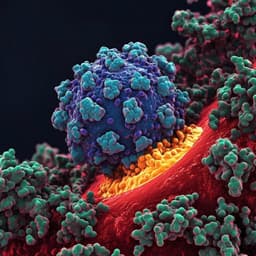
The spike gene is a major determinant for the SARS-CoV-2 Omicron-BA.1 phenotype
M. P. Alves, C. G. Benaraï, et al.
SARS-CoV-2 evolution has been characterized by successive waves of variants of concern (VOCs), notably Alpha, Delta, and Omicron. Delta harbors mutations such as L452R (associated with immune escape) and P681R (associated with enhanced transmission). Omicron-BA.1 contains an extensive set of mutations (up to 50 total; 34 in spike, 15 in the RBD), including a defining insertion ins214EPE whose contribution to fitness is unclear. Omicron-BA.1 exhibits marked antibody escape (up to 40-fold greater than ancestral and pre-Omicron variants) and rapidly became globally predominant by January 2022 before being replaced by BA.2 and other sublineages. It remains unresolved whether BA.1’s rapid spread relative to Delta is driven primarily by intrinsic fitness/transmission or immune escape in vaccinated populations. The genetic determinants of the Omicron-BA.1 phenotype also remain poorly defined. Given concurrent circulation of multiple VOCs and reports of recombination, there is an urgent need to dissect fitness and immune escape of VOCs using advanced cell culture and animal models (hamsters, ferrets, and human ACE2-expressing mice) to clarify the role of spike in BA.1’s phenotype.
Prior work has identified key Delta spike mutations: L452R enhances immune escape and infectivity; P681R increases fitness and transmission. Spike D614G was shown to enhance replication and transmission. Omicron-BA.1 carries numerous spike mutations, especially in the RBD, enabling substantial neutralization escape versus vaccine-elicited antibodies. Animal models differ in susceptibility and disease severity: Syrian hamsters and hACE2-expressing mice typically show severe disease, whereas ferrets display subclinical infection despite efficient replication with earlier variants. Ex vivo studies reported altered replication of Omicron in human bronchus and lung tissue. The role of spike-specific changes, including insertions such as ins214EPE, in BA.1’s fitness and tropism remained uncertain, necessitating direct comparative studies across primary human airway cultures and multiple animal hosts.
Biosafety: All infectious SARS-CoV-2 work was conducted in BSL-3 facilities at the Institute of Virology and Immunology (Switzerland) and Friedrich-Loeffler-Institut (Germany) under approved SOPs and ethical oversight.
Cells: VeroE6/TMPRSS2, Vero cell derivatives, BHK-21 cells expressing SARS-CoV-2 S, and Calu-3 cells were maintained under standard conditions with supplemented media. Primary well-differentiated human nasal (hNEC) and bronchial (hBEC) epithelial cultures were generated at air–liquid interface following established protocols.
Viruses: VOC isolates (Alpha, Delta AY.127, Omicron-BA.1) and recombinant clones differing in spike sequence were used, with stocks propagated on appropriate cell lines. Sequences were verified.
Primary airway infection and kinetics: hNECs (33 °C) and hBECs (37 °C) were infected apically with defined doses (e.g., 100 TCID50; or 10^6 TCID50 for some experiments). Apical washes were collected at set intervals up to 96 hpi and titrated by TCID50 on VeroE6/TMPRSS2. Competition assays used 1:1 GE mixtures (6 × 10^7 GE each virus) with apical infections and sampling at 2, 4, and 6 dpi, followed by sequencing and RT-qPCR to quantify variant proportions.
Animal studies: Syrian hamsters were intranasally inoculated with mixtures of VOCs (e.g., Delta vs Omicron-BA.1), followed by sequential co-housing of naïve contacts (Contact I and II). Nasal washings were taken daily; donors were sampled for organs at 4 dpi. Ferret competition and single-infection studies used intranasal inoculation with mixtures or single VOCs, daily nasal swabbing, and contact exposure via co-housing or shared air, with sampling up to 21 dpi and organ analysis at defined time points. hACE2 knock-in (hACE2-KI) mice were intranasally inoculated (e.g., 10^3 TCID50/mouse) with Delta, Omicron-BA.1, or 1:1 mixtures of VOC isolates or spike-differing recombinant clones to assess replication, tissue distribution (URT, LRT, brain/olfactory bulb), pathology, and competitive fitness. K18-hACE2 mice (female 7–15 weeks) received a single intramuscular dose of mRNA SpikeVax (Moderna), then were challenged intranasally with SARS-CoV-2 clones (index, Delta spike clone, Omicron-BA.1 spike clone). Tissues (nose, lung, brain) and oropharyngeal swabs were collected at 2 and 6 dpi to assess viral RNA and infectious titers.
Molecular analyses: Variant-specific RT-qPCR quantified genome copies and variant ratios. TCID50 assays on VeroE6/TMPRSS2 quantified infectious virus. Nanopore sequencing (Oxford Nanopore MinION) followed ARTIC v3/Midnight primer schemes with Omicron-specific primers to ensure balanced amplification. Additional library preparation and Illumina-based sequencing were used for confirmation.
Pathology and IHC: Formalin-fixed paraffin-embedded lungs and brains from mice were H&E-stained and scored; IHC for SARS-CoV nucleocapsid antigen was performed using standard protocols.
Serology: Live virus neutralization tests (VNT100) and multispecies RBD ELISA assessed neutralizing capacity and seroreactivity.
Statistics: GraphPad Prism 8; comparisons included two-way ANOVA with Tukey’s multiple-comparison test for in vitro assays and unpaired two-tailed Student’s t-test for selected in vivo endpoints, with conventional significance thresholds.
Ethics: All animal and human tissue-related procedures were approved by relevant institutional and governmental committees and conducted under applicable regulations.
- Omicron-BA.1 spike-driven advantage in nasal epithelium: In well-differentiated human nasal epithelial cultures, Omicron-BA.1 (isolate and spike clone) showed accelerated replication and greater replicative fitness compared to pre-Omicron spike genes, whereas replication in bronchial epithelial cells was limited relative to Delta.
- Syrian hamsters: In co-infection/competition experiments, Delta consistently dominated over Omicron-BA.1 in nasal wash and organ samples of contact animals. Donor animals shed high viral genome loads, with nasal wash peaks >10^7 GE/ml at 5–6 dpi; two of three contact animals were infected by 6 days post-contact with loads up to 10^6 GE/ml. Delta was detected throughout URT and LRT at 4 dpi with highest loads in the nasal cavity (~10^6 GE/ml). Neutralizing antibody responses were primarily directed against Delta, reaching serum dilutions up to 1:1024.
- Ferrets: In co-inoculated donors and contacts, only Delta was detected in nasal washings beginning at 1 dpi with levels up to 10^7 GE/ml; 5/6 contact ferrets shed >10^6 GE/ml starting at 2 dpi, with shedding up to 12 days. Omicron-BA.1 was not detected in donors or contacts, indicating abortive infection and failure of transmission for BA.1 in ferrets under these conditions.
- hACE2-KI mice: Delta caused greater body weight loss (noted at 4 dpi), higher viral RNA loads and infectious titers in URT, LRT, and olfactory bulb, and higher lung pathology scores than Omicron-BA.1. In direct competition, Delta dominated Omicron-BA.1 in both URT and LRT. When comparing recombinant clones differing only by spike sequence, the Delta spike clone fully dominated the Omicron-BA.1 spike clone, indicating spike-mediated fitness differences.
- K18-hACE2 mice and vaccination: The Omicron-BA.1 spike conferred a less virulent phenotype (lower infectious titers in nose, lung, brain of unvaccinated mice versus Delta) but a higher degree of immune evasion. In vaccinated mice, infectious titers for Omicron-BA.1 in the nose at 2 and 6 dpi were comparable to unvaccinated animals, while in the lung and brain infectious virus was reduced or undetectable, demonstrating vaccine-mediated protection primarily in the lower respiratory tract and CNS. Vaccination reduced replication and pathogenesis for index and Delta spike clones more effectively than for the Omicron-BA.1 spike clone.
- Statistical signals: In vitro comparisons (e.g., plaque size/replication kinetics) reported significance by two-way ANOVA with Tukey’s adjustment (p < 0.05 to p < 0.0001) for Omicron vs controls; in vivo mouse comparisons used unpaired two-tailed t-tests with conventional significance thresholds. Overall, data support that BA.1’s spike enhances early replication in the nasal epithelium and mediates immune evasion, while Delta’s spike drives superior fitness and pathogenicity in lower airways and across several animal models.
The study addressed whether the Omicron-BA.1 spike underlies the phenotypic traits that enabled BA.1 to overtake Delta, distinguishing between intrinsic fitness/transmissibility and immune escape. Findings show that the BA.1 spike confers a replication advantage specifically in the human nasal epithelium, consistent with enhanced upper-airway tropism, while exhibiting limited replication in bronchial epithelium compared to Delta. In vivo, Delta outcompeted BA.1 in hamsters and hACE2-KI mice and was the only variant to productively infect and transmit among ferrets, indicating that Delta retains superior fitness across these animal hosts, especially for LRT replication and pathogenesis. Spike-only recombinant competitions confirmed that these differences are spike-mediated. In K18-hACE2 mice, the BA.1 spike produced reduced pathogenicity but showed substantial vaccine escape in the URT: vaccination reduced LRT and brain infection but had limited impact on nasal infection by the BA.1 spike clone. Together, these results indicate that BA.1’s success in humans likely reflects a combination of spike-mediated immune evasion and preferential upper-airway replication rather than broadly increased intrinsic fitness across hosts or lower-airway tissues. These insights underscore the central role of spike mutations in shaping tissue tropism, host range, and vaccine effectiveness.
This work demonstrates that the SARS-CoV-2 Omicron-BA.1 spike gene is a major determinant of its phenotype: it enhances early replication in the nasal epithelium, reduces lower-airway replication and pathogenicity relative to Delta, and mediates significant immune evasion, particularly in the upper respiratory tract. Delta’s spike, by contrast, confers higher fitness and pathogenicity in multiple animal models and dominates BA.1 in direct competition. These findings help explain BA.1’s rapid spread in humans as driven by spike-dependent immune escape and URT tropism rather than generalized increases in intrinsic fitness. Future studies should map individual BA.1 spike mutations to specific phenotypic effects, assess later Omicron sublineages and recombinants, and further dissect tissue tropism and transmission dynamics in human-relevant systems.
Related Publications
Explore these studies to deepen your understanding of the subject.