
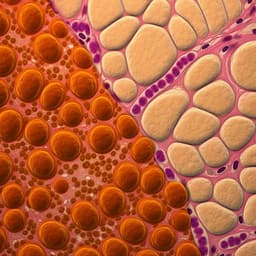
The expression signatures in liver and adipose tissue from obese Göttingen Minipigs reveal a predisposition for healthy fat accumulation
S. Cirera, E. Taşöz, et al.
Explore these studies to deepen your understanding
Adjacent work that informs or extends this paper's methodology and findings.

MyD88 determines the protective effects of fish oil and perilla oil against metabolic disorders and inflammation in adipose tissue from mice fed a high-fat diet
F. Wang, M. Hu, et al.
Maternal high-fat diet programs white and brown adipose tissue lipidome and transcriptome in offspring in a sex- and tissue-dependent manner in mice
C. Savva, L. A. Helguero, et al.
Impact of baseline adipose tissue characteristics on change in adipose tissue volume during a low calorie diet in people with obesity—results from the LION study
D. Junker, M. Wu, et al.
Commensal *Hafnia alvei* strain reduces food intake and fat mass in obese mice—a new potential probiotic for appetite and body weight management
R. Legrand, N. Lucas, et al.


