
Biology
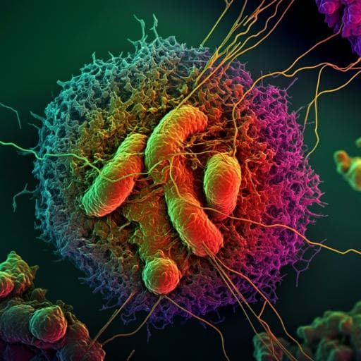
Single-particle cryo-EM structures from IDPC-STEM at near-atomic resolution
I. Lazić, M. Wirix, et al.
This groundbreaking study showcases the potential of integrated differential phase contrast scanning transmission electron microscopy (IDPC-STEM) for high-resolution structure determination in cryo-electron microscopy. By examining keyhole limpet hemocyanin and tobacco mosaic virus, the researchers achieved near-atomic resolution, establishing IDPC-STEM as a powerful tool for biological macromolecule analysis. This research was conducted by Ivan Lazić, Maarten Wirix, Max Leo Leidl, Felix de Haas, Daniel Mann, Maximilian Beckers, Evgeniya V. Pechnikova, Knut Müller-Caspary, Ricardo Egoavil, Eric G. T. Bosch, and Carsten Sachse.
~3 min • Beginner • English
Introduction
Scanning transmission electron microscopy (STEM) is widely used for materials characterization and, in advanced modes such as ptychography, can achieve sub-Å resolution. Among STEM modalities, integrated differential phase contrast (IDPC-STEM) has been successfully applied to crystalline and amorphous materials, including beam-sensitive systems, enabling high-contrast, in-focus imaging at low doses. In biological EM, cryo-EM typically uses conventional TEM (CTEM) at significant underfocus to boost low-frequency contrast and combines many micrographs to obtain near-atomic 3D structures. While cryo-STEM has been used for tomography of thick cells and for locating metals in proteins, complete single-particle 3D reconstructions of biological macromolecules at near-atomic resolution using STEM images had not been demonstrated. This study investigates whether IDPC-STEM can be applied to vitrified biological single-particle specimens to yield high-resolution micrographs suitable for 3D reconstruction. The authors apply IDPC-STEM to keyhole limpet hemocyanin (KLH) and tobacco mosaic virus (TMV), aiming to achieve subnanometer and near-atomic resolutions and to benchmark performance against CTEM datasets.
Literature Review
Prior STEM work has demonstrated imaging at better than 0.5 Å resolution and successful visualization of diverse materials, including dose-sensitive zeolites and metal–organic frameworks, often with low electron doses. IDPC-STEM has been shown to visualize both heavy and light elements and to provide dose-efficient, high-contrast images for thick biological sections, enabling direct interpretation without defocus and CTF correction. In biological cryo-EM, CTEM advancements (the 'resolution revolution') have routinely delivered near-atomic structures; benchmark specimens like TMV and apoferritin have reached 2.0 Å and 1.2 Å, respectively, leveraging direct electron detectors and improved image processing. Previous cryo-STEM applications include tomography of vitrified cells and low-resolution imaging of virus-like particles by ptychography, but not single-particle near-atomic 3D reconstructions. Limitations of other STEM modes for low-dose biological imaging are noted: ABF-STEM requires defocus to transfer low frequencies and collects only a fraction of electrons, and ADF-STEM, while in-focus and positive CTF, is dose-inefficient and suppresses light elements due to intensity dependence on the square of electrostatic potential. IDPC-STEM, by contrast, uses all relevant electrons, integrates to suppress noise, features a favorable, non-oscillatory CTF without zero crossings, and produces images proportional to the electrostatic potential, suggesting suitability for cryo-EM.
Methodology
Specimen preparation: Quantifoil grids (R2/2, 200 mesh) were glow-discharged (45 s, air). For TMV, 3.5 µl of ~90 mg/ml (~2.5 µM) solution was applied, blotted in a Vitrobot Mark IV at 4 °C/100% RH with high blot force (+10) and 10 s blot time to obtain thin ice across 2 µm holes; grids were plunge-frozen and stored in liquid nitrogen. For KLH, a 10 mg/ml (~2.5 µM) solution was used with blot force −10 and 6 s blot time.
Microscope and detectors: A Thermo Fisher Scientific Titan Krios G4 at 300 kV with X-FEG, three-condenser lens system, C-TWIN objective (11 mm pole piece, Cs 2.7 mm), Panther segmented STEM detector (four-quadrant IDPC readout), Ceta camera and Falcon 4 DED was used. Convergence semiangles (CSA) were set via C2 apertures (20/50 µm) and C2/C3 lens ratios. Precise COM determination included beam shift/tilt pivot alignment and descan alignment. CSAs were calibrated using Au cross-grating rings and bright-field disk radii on the Ceta camera.
IDPC-STEM acquisition: The four-quadrant Panther detector recorded IDPC signals. The beam size on the detector was set to cover >2/3 of the detector diameter; the central hole radius was <1/4 of the beam size. A simple line-by-line scan grid was used; the scan direction offset relative to detector quadrants was measured and compensated when computing DPC vector components prior to integration. Camera length for each CSA ensured the bright-field disk radius was ~4× the central hole radius. For CSA ≥4.5 mrad, Au rings in IDPC and ADF modes confirmed attainable resolution. The beam current was fixed at 4 pA; dwell time was tuned to reach a total electron dose of 35 e−/Ų (TMV) or ~40 e−/Ų (KLH example). Typical 4,096×4,096 acquisitions took 13.2 s (0.75 Å pixel) to 67.9 s (1.70 Å pixel), with ~10 s rest between frames.
Imaging conditions and scanning: Unlike CTEM flood-beam, a convergent probe scans the specimen spot-by-spot; for focus determination, CBED (Ronchigram) flatness was used. The CSA controls apparent resolution and depth of focus; in-focus imaging provides maximal contrast (no defocus used). Example at CSA 2.0 mrad: probe effective diameter 4.9 Å, 2.4 Å scan step (50% overlap), 4 µs dwell, 4,096×4,096 pixels, 68 s total, ~983 nm FOV. Optical resolution verification on 50 nm Au-on-carbon (CSA 4.5 mrad, 350 e−/Ų) showed the 2.3 Å gold ring, near theoretical 2.2 Å for that CSA, and absence of CTEM Thon rings; a fourfold star pattern at low frequencies reflected the IDPC-STEM CTF.
Data acquisition: KLH—760 micrographs at CSA 2.0 mrad and 687 at CSA 3.5 mrad (300 kV; example pixel sizes 1.5 Å for raw, 1.2 Å for CSA 3.5 mrad; doses ~35–40 e−/Ų). TMV—sets at CSAs 2.0, 3.0, 3.5, 4.0, 4.5 mrad (300 kV), with pixel sizes matched to CSA to avoid aliasing (e.g., 0.98 Å at 4.0 mrad), 4,096×4,096 pixels, ~35 e−/Ų. Images were preprocessed with a Gaussian high-pass filter (FWHM 251 Å) and not contrast-inverted.
Image processing: KLH—CryoSPARC v3.3.1. CSA 2.0 mrad: 9,750 particles extracted; 2D classification; ab initio 3D model (batch size 1,000). CSA 3.5 mrad: ~500 manually picked for templates; 120,727 picked; after 2D classification (batch size 200, 200 iterations), 18,597 retained; refinement with initial low-pass filtered (60 Å) model and D5 symmetry; non-uniform local refinement yielded 6.5 Å (FSC 0.143) / 6.8 Å (FDR-FSC) final map. Map sharpened by Guinier B factor (~523 Ų). PDB 4BED docked.
TMV—Segment picking in EMAN2; averaged power spectra computed from in-plane rotated segments; 1D helical profiles used to calibrate pixel size. RELION v3.1 helical workflow: 1–2 rounds 2D classification; no CTF estimation/correction (in-focus, positive CTF). CTEM reference subsets (EMPIAR-10305, EMPIAR-10021) reprocessed analogously with motion correction and limited dose for comparability. 3D classification with featureless cylinder; refinement; mask of central 30% in z; postprocessing and local resolution estimation. FSC at 0.143 and mask-free FDR-FSC used. Atomic models PDB 6SAG (EMPIAR-10305) and 4UDV (EMPIAR-10021 and IDPC-STEM maps) rigid-body fitted; visualization with UCSF Chimera/ChimeraX and Coot.
Simulations: Multislice STEM simulations (extended for IDPC-STEM) examined SNR vs CSA (smaller CSA gives higher SNR for fixed dose) and aliasing at high CSA with insufficient sampling; acquisition parameters set to meet Nyquist for CSA-dependent resolution as per Table 1.
Key Findings
• IDPC-STEM micrographs exhibit continuous, positive contrast transfer across spatial frequencies without CTF zero crossings; low spatial frequencies are strong relative to defocused CTEM images.
• Optical performance verified on Au-on-carbon: 2.3 Å ring resolved at CSA 4.5 mrad (theoretical limit 2.2 Å), confirming near-limit resolution transfer.
• KLH single-particle reconstruction from IDPC-STEM achieved subnanometer resolution: 6.5 Å (FSC 0.143; 6.8 Å FDR-FSC) with D5 symmetry using 18,597 particles (CSA 3.5 mrad). PDB 4BED docked with agreement to expected secondary structure.
• TMV helical reconstructions from IDPC-STEM at multiple CSAs reached near-atomic resolution: 6.3 Å (2.0 mrad), 4.3 Å (3.0 mrad), 3.9 Å (3.5 mrad), 3.5 Å (4.0 mrad; best), 3.7 Å (4.5 mrad) by FSC 0.143; corresponding FDR-FSC: 5.9, 4.1, 3.4, 3.2, 3.3 Å. The 4.0 mrad map at 3.5 Å shows side-chain densities for large/aromatic/positively charged residues and well-defined backbone; RNA density visible. Negatively charged side-chain densities are weak/absent, similar to CTEM.
• Helical layer-line analysis: the 1D profile decays faster for IDPC-STEM than defocused CTEM; the second/first layer-line intensity ratio increases with CSA (0.2 at 2.0 mrad to 0.5 at 4.5 mrad), indicating improved high-frequency transfer at higher CSA but still below latest CTEM (ratio ~1.9 for EMPIAR-10305).
• Guinier B factors for TMV maps (IDPC-STEM): 492 (2.0 mrad), 155 (3.0 mrad), 105 (3.5 mrad), 132 (4.0 mrad), 126 Ų (4.5 mrad); global B factor from particle-subset analysis ~93 Ų (IDPC-STEM 4.0 mrad TMV), better than 2015 CTEM (Falcon II) and worse than 2019 CTEM (K2).
• Acquisition feasibility: Without motion correction or advanced scan strategies, simple line scanning and four-quadrant detection yield near-atomic TMV structures; observed Fourier transforms show characteristic TMV layer lines and no CTF zeros; a vertical meridian streak reflects known STEM scanning effects.
• Depth-of-focus considerations: At 300 kV, CSAs up to 4.5 mrad (depth of focus 194 nm) produce complete projections for typical ice thicknesses (10–60 nm).
Discussion
The findings demonstrate that IDPC-STEM can produce high-contrast, in-focus images of vitrified macromolecules with continuous information transfer to high resolution, suitable for single-particle and helical cryo-EM reconstructions at near-atomic levels. Compared to ABF- and ADF-STEM, IDPC-STEM optimally uses available electrons, suppresses noise during integration, and avoids CTF oscillations and zero crossings; images are proportional to the electrostatic potential, preserving light elements. Relative to CTEM, the scanning delivery of electrons may allow energy dissipation between illuminated pixels and offers flexibility in scan patterns that could mitigate radiation damage and motion. The observed performance depends critically on CSA, which sets both lateral resolution and depth of focus; increasing CSA improves lateral resolution but reduces depth of focus and, at sufficiently high values and with aberrations, can introduce CTF zero crossings, motivating aberration correction for higher CSAs. For typical cryo-EM ice thicknesses tested, CSAs up to 4.5 mrad yielded complete projections and high-resolution reconstructions. While IDPC-STEM helical profiles decay faster than defocused CTEM, near-atomic reconstructions comparable to a 2015 CTEM dataset were achieved and were outperformed by a 2019 CTEM dataset, as reflected in B factors and resolution. The approach currently lacks frame-based motion correction and uses relatively slow, manual acquisition; however, detector capabilities permit much faster scan speeds and automation, which are expected to reduce acquisition times to below a second per frame and enable large datasets akin to CTEM. The technique also opens avenues for imaging thicker specimens via optical sectioning, applying dynamic focusing to counter preferred orientations, performing cryo-electron tomography with non-oscillatory CTF, and integrating spectroscopic mapping for correlative analyses.
Conclusion
This work establishes the principal applicability of IDPC-STEM to cryo-vitrified biological single-particle specimens for high-quality imaging and 3D structure determination at near-atomic resolution. Subnanometer KLH (6.5 Å) and near-atomic TMV (to 3.5 Å) structures were reconstructed from IDPC-STEM micrographs acquired in focus without CTF correction. The results validate that appropriate combinations of CSA and sampling achieve resolutions within theoretical limits and yield map features comparable to CTEM at similar resolutions. Future research should focus on: automation and high-speed scanning to scale data acquisition; movie-mode frame recording and motion correction to further improve map quality; use of aberration-corrected probes for higher CSAs; exploration of alternative scan strategies to mitigate radiation damage; application to thicker specimens via optical sectioning and to cryo-electron tomography; deployment in complex cellular contexts where improved low-frequency contrast is advantageous; and integration with spectroscopic techniques for correlative structural and compositional analyses.
Limitations
Current IDPC-STEM datasets were modest in size and acquired without automation, limiting throughput compared to CTEM. Scan-induced artifacts (e.g., vertical streaks) and potential local motion can introduce distortions; no motion correction was applied. Helical power spectra from IDPC-STEM decay faster than from defocused CTEM, and achieved resolutions, while near-atomic, did not reach the best CTEM benchmarks. The trade-off between CSA and depth of focus imposes constraints; at higher CSAs and in the presence of aberrations, CTF zero crossings can appear, necessitating aberration correction. Signal-to-noise decreases with increasing CSA for fixed dose, and aliasing can occur if sampling does not match optical resolution, requiring careful choice of pixel size. Densities for negatively charged side chains remain weak due to radiation damage, similar to CTEM.
Related Publications
Explore these studies to deepen your understanding of the subject.


