logo
ResearchBunny Logo
Single-molecule amplification-free multiplexed detection of circulating microRNA cancer biomarkers from serum

Medicine and Health

Single-molecule amplification-free multiplexed detection of circulating microRNA cancer biomarkers from serum

S. Cai, T. Pataillot-meakin, et al.

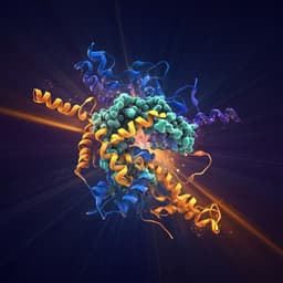
This groundbreaking research by Shenglin Cai and colleagues introduces size-encoded molecular probes that allow for electro-optical nanopore sensing of microRNAs in unprocessed human serum. Achieving remarkable sensitivity and specificity, this innovative approach may revolutionize cancer diagnostics.

00:00
00:00
~3 min • Beginner • English
Introduction
Prostate cancer (PCa) is the second most prevalent male cancer worldwide and current monitoring relies heavily on serum PSA testing, which suffers from low specificity and sensitivity, temporal variability, and delays in reflecting treatment effects. Liquid biopsies offer access to circulating biomarkers (cells, exosomes, nucleic acids) that could improve monitoring, but translation has been limited by analytical challenges in detecting scarce targets directly from biofluids. Circulating miRNAs, particularly miR-141-3p and miR-375-3p, are dysregulated in PCa and hold promise as minimally invasive biomarkers; however, conventional workflows require RNA extraction and PCR amplification, introducing bias and limiting quantitation. Existing single-molecule fluorescence methods face multiplexing constraints, and label-free nanopore sensors to date have lacked adequate sensitivity and multiplexing for rare, highly homologous miRNAs. The study aims to develop and validate an amplification- and extraction-free, multiplexed, single-molecule platform capable of ultrasensitive and sequence-specific detection of multiple circulating miRNAs directly in human serum, and to evaluate its clinical utility in distinguishing PCa disease states.
Literature Review
Prior approaches to circulating miRNA analysis include bead- or electrode-bound capture, microfluidic platforms, and single-molecule fluorescence microscopy. These methods often require expensive consumables, larger sample volumes, or suffer from limited multiplexing due to fluorophore constraints. Label-free nanopore sensing has emerged for single-molecule detection, including miRNAs, but has been constrained by limited multiplexing capacity and insufficient sensitivity/selectivity for clinically relevant, rare targets. Conventional RT-qPCR, microarrays, and RNA-Seq provide sensitivity but require extraction and amplification, introducing pre-analytical variability and potential bias, and are resource- and time-intensive. Recent nanopore methods achieved mainly nM to pM sensitivity, typically necessitating pre-enrichment for biofluids, underscoring the need for improved techniques.
Methodology
The authors developed an electro-optical nanopore platform integrating synchronized electrical nanopore sensing using quartz nanopipettes with confocal fluorescence detection. Molecular probes consisted of long dsDNA carriers (lambda DNA fragments) barcoded by length (5.6, 10, and 38.5 kbp) and grafted with fluorophore/quencher-labelled molecular beacons (MBs) specific to target miRNAs. Upon hybridization to a complementary miRNA, the MB hairpin opens, restoring fluorescence. The electrical signal reports carrier length (barcode) via dwell time and peak current area, while the optical channel reports target binding (yes/no). Multiplexing is achieved by assigning distinct carrier lengths to different MBs. Preparation: Lambda DNA was enzymatically digested (Apa I for 10 and 38.5 kbp; BstE II for 5.6 kbp); MBs specific to miR-375-3p, miR-141-3p, let-7a/let-7b, and miR-21 were hybridized to 12-base sticky overhangs; unbound components were removed via ultrafiltration or gel extraction. Nanopipettes (~22 ± 3 nm pores) were fabricated by laser pulling from quartz capillaries. Electro-optical setup: A 488 nm confocal microscope (APD detection) was aligned to the nanopipette tip inside a Faraday cage; electrical signals were recorded with a patch-clamp amplifier at 100 kHz sampling (10 kHz filter). Synchronization used dual NI DAQ cards and LabVIEW. Event detection thresholds were set at multiples of baseline noise (electrical 7σ, optical 5σ), and coincident events were identified by timestamp cross-correlation. Experimental conditions: Symmetric buffer (100 mM KCl, 5 mM MgCl2, 10 mM Tris-HCl, 1 mM EDTA, pH 8.0) at −300 mV was used for initial characterization. To enhance capture rates and sensitivity, an asymmetric salt gradient (cis 40 mM KCl; trans 400 mM KCl) was employed with lower probe concentrations (1 pM). Synchronization ratio S was defined as synchronised optical-electrical events divided by total electrical events per barcode, used for quantitation. Calibration: Titrations with synthetic miRNAs (0.1–100 pM) at fixed probe concentration determined linear ranges and LODs; capture rate improvement with salt gradient was quantified. Specificity tests used highly homologous family members (let-7a vs let-7f; miR-141-3p vs miR-200a-3p) and scrambled controls. Serum handling: Human serum was mixed with probes at optimized dilution (1:10 serum:buffer for stability/sensitivity balance), incubated 2 h, and ~1 µl loaded into the nanopipette; measurements performed under salt gradient at −300 mV. Comparative analyses included measurements in RNA extracts (from 0.5 ml serum using Monarch kit) and RT-qPCR (Exiqon LNA system). Data analysis used custom Matlab apps for event classification by barcode (dwell time and peak area), S computation, concentration estimation via calibration curves, and statistical testing (two-tailed Student’s t-tests; Pearson correlations).
Key Findings
- Distinguishable electrical barcodes: Three DNA carrier lengths (5.6, 10, 38.5 kbp) showed distinct current transients with dwell times of 0.45 ± 0.19 ms, 1.1 ± 0.3 ms, and 5.0 ± 4.1 ms and peak current areas of 9.3 ± 5.5 fAs, 26.9 ± 7.1 fAs, and 189.7 ± 123.5 fAs (mean ± s.d.), enabling multiplexing. - Multiplexed detection with synthetic targets: For Probe-375 (10 kbp) and Probe-141 (38.5 kbp), baseline synchronisation ratios S without targets were ~0.8–0.9%. With target present, S rose to 76.1 ± 7.6% (miR-375-3p) and 80.5 ± 9.2% (miR-141-3p), with negligible cross-reactivity; discrimination accuracy was 98.8% at picomolar concentrations. In a 3-plex (let-7a/miR-375-3p/miR-141-3p), S increased from ~1–2% baseline to 55.5 ± 9.5%, 75.9 ± 9.2%, and 78.8 ± 7.6%, respectively. - Sensitivity: Calibration yielded linear S vs concentration from 0.1–10 pM (R² ≥ 0.996). Initial LODs (3σ) were 0.13 pM (miR-375-3p) and 0.10 pM (miR-141-3p). Using a 40/400 mM KCl gradient and 1 pM probes increased capture rate ~8× (0.15 ± 0.03 s⁻¹ to 1.19 ± 0.12 s⁻¹) and improved LODs to 8 fM (miR-375-3p) and 5 fM (miR-141-3p). Benchmarking showed markedly worse LODs for conventional single-molecule confocal and bulk fluorescence (e.g., 0.67 pM/3.2 nM for miR-375-3p; 1.01 pM/5.1 nM for miR-141-3p). - Single-nucleotide specificity: Perfectly matched targets produced high S (~76–79%), whereas single mismatch (let-7f) and double mismatch (miR-200a-3p) yielded significantly lower S (10.6 ± 5.3% and 4.6 ± 2.6%; P < 0.0001). Scrambled controls matched blank levels (~0.9%). - Direct detection in serum: Nanopipette confinement reduced autofluorescence up to 50× in 1:2 serum:buffer, enabling single-molecule detection. Electrical baselines were stable for serum dilutions ≥1:5; 1:10 selected as optimal. - Clinical PCa cohorts (direct serum, n=5 active; n=5 remission): Active disease showed higher S for both miRNAs; calculated average relative expression (normalized to remission = 1) increased 12.5 ± 1.6× (miR-141-3p) and 4.2 ± 0.9× (miR-375-3p), with P < 0.0001. Sensitivity and specificity were 100% for distinguishing active vs remission using either miRNA. - Extracted RNA and RT-qPCR comparisons: In extracts, active vs remission differences were smaller (3.1× miR-141-3p, P=0.0166; 2.8× miR-375-3p, P=0.0488) with lower sensitivities (80% and 60% at 100% specificity). RT-qPCR showed non-significant trends (miR-141-3p P=0.724; miR-375-3p P=0.079). Correlations: serum vs extract concentrations showed Pearson r=0.7038 (miR-141-3p, P=0.0231) and r=0.8217 (miR-375-3p, P=0.0035). Extract nanopore vs RT-qPCR Ct: r=−0.6346 (miR-141-3p, P=0.0485) and r=−0.9436 (miR-375-3p, P=0.0001). - Staging panel (3-plex: miR-141-3p, miR-375-3p, let-7b): In cohorts (remission n=7; advanced localised n=3; metastatic n=3), miR-141-3p and miR-375-3p were progressively upregulated from remission to localised (P<0.05) to metastatic (P<0.05), while let-7b was downregulated in advanced disease; localised vs metastatic differentiation achieved with moderate significance (P<0.05). - Practical metrics: Sample volume as low as ~0.1 µl; quantitation down to ~5 zeptomoles (~10^3 copies). Estimated assay turnaround ~80 min for multi-target profiling, faster than RT-qPCR, microarrays, or RNA-Seq.
Discussion
The platform directly addresses the need for sensitive, specific, and multiplexed detection of scarce circulating miRNAs without extraction or amplification. By coupling electrical barcoding of DNA carriers with optical readout of target binding, the method achieves femtomolar sensitivity in serum, surpassing prior nanopore and fluorescence approaches and approaching RT-qPCR performance while avoiding its biases. Single-nucleotide specificity mitigates false positives among highly homologous miRNA families, crucial for clinical specificity. Direct analysis from serum reduces pre-analytical variability and preserves native distributions of circulating miRNAs, potentially capturing clinically relevant dynamics more accurately than extract-based methods. The strong discrimination between active PCa and remission, and the ability to stratify disease stage using a three-miRNA panel, demonstrate clinical relevance. The approach is scalable in multiplexing via additional carrier lengths and potentially multiple fluorophores, and the small sample volume and rapid turnaround support point-of-care potential. Correlations with extracts and RT-qPCR highlight differences due to extraction losses, compartmentalization (free vs vesicle-encapsulated), and amplification variability, further underscoring the value of direct, extraction-free measurements.
Conclusion
This work introduces an amplification- and extraction-free electro-optical nanopore method for multiplexed, single-molecule detection of circulating miRNAs directly in human serum. Using size-encoded DNA carrier–molecular beacon probes, the platform achieves femtomolar sensitivity, single-base mismatch specificity, and requires only ~0.1 µl of sample. It discriminated active prostate cancer from remission with 100% sensitivity and specificity using either miR-141-3p or miR-375-3p, and a three-miRNA panel enabled differentiation of disease stages. The method outperforms conventional fluorescence assays and compares favorably to RT-qPCR while avoiding pre-analytical and amplification biases. Future directions include expanding multiplexing capacity (more carrier barcodes and/or multicolor fluorophores), improving capture via nanopore engineering or dielectrophoresis, integrating exosome/vesicle lysis for comprehensive miRNA profiling, validating in larger cohorts and whole blood, and extending to other biomarkers (RNAs, cfDNA, proteins) for multi-omic, multi-disease screening.
Limitations
- Small clinical cohorts (active n=5, remission n=5; staging cohorts localised n=3, metastatic n=3) limit generalizability; findings require validation in larger, independent populations. - Electrical stability required serum dilution (optimal 1:10), which may limit sensitivity for very low abundance targets; higher serum fractions increased autofluorescence and reduced electrical stability. - Detection sensitivity at the lowest concentrations is limited by capture frequency; improvements rely on salt gradients or advanced concentration strategies (e.g., dielectrophoresis). - Multiplexing capacity is constrained by electrical resolution of carrier lengths; authors estimate ~10 targets feasible with λ-DNA unless further pore/measurement optimizations are implemented. - Direct serum measurements may preferentially detect freely circulating/protein-bound miRNAs and underrepresent vesicle-encapsulated species; differences from extract-based methods may reflect compartmentalization. - Platform requires precise electro-optical alignment and specialized instrumentation, which may affect reproducibility without standardization.
Listen, Learn & Level Up
Over 10,000 hours of research content in 25+ fields, available in 12+ languages.
No more digging through PDFs, just hit play and absorb the world's latest research in your language, on your time.
listen to research audio papers with researchbunny