Medicine and Health
Single-cell guided prenatal derivation of primary fetal epithelial organoids from human amniotic and tracheal fluids
M. F. M. Gerli, G. Calà, et al.
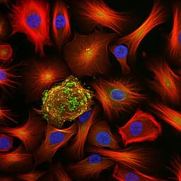
This groundbreaking research led by Mattia Francesco Maria Gerli and colleagues unveils a novel method for isolating viable epithelial stem cells from human amniotic fluid. These cells demonstrated the ability to form organoids that mimic the small intestine, kidney tubules, and lungs, including those with congenital diaphragmatic hernia features. This innovative approach opens new avenues for organoid derivation during pregnancy, paving the way for personalized therapies and regenerative medicine.
~3 min • Beginner • English
Introduction
The study addresses the challenge of generating autologous, patient-specific models of developing human tissues during ongoing pregnancy to improve prenatal diagnosis, severity prediction, counseling, and therapy selection for congenital anomalies. Despite advances in genetic and imaging diagnostics, predicting disease severity is difficult and limits personalized prenatal interventions. Existing primary fetal organoids are usually derived destructively from postmortem fetal tissues, restricted by ethical/legal constraints and gestational age (typically up to 20–22 weeks), and iPSC-based organoids require extensive manipulation and long differentiation timelines, reducing fidelity and prenatal applicability. The authors hypothesize that epithelial stem/progenitor cells shed into fetal fluids (amniotic fluid and tracheal fluid) can be isolated and expanded into primary fetal organoids representing their tissues of origin (gastrointestinal, kidney, lung), enabling timely, minimally invasive, autologous prenatal disease modeling (including congenital diaphragmatic hernia) and potential therapeutic testing.
Literature Review
The paper contrasts iPSC-derived organoids (including those generated from reprogrammed AF cells) that resemble fetal-like tissues but require lengthy, complex differentiation and manipulation, with primary organoids derived from minimally manipulated, discarded postnatal samples (urine, menstrual flow, PAP smear, BAL). Prior prenatal primary organoids relied on fetal tissues obtained postmortem, limited by ethics and gestational windows, impeding prenatal modeling and diagnostics. AF is known to contain heterogeneous cells from multiple fetal organs and mesenchymal/hematopoietic niches, yet epithelial identities and organoid-forming potential had not been comprehensively mapped. This study builds on single-cell atlases of fetal tissues but addresses gaps at later gestational stages and cells outside native tissue context.
Methodology
- Ethics: Fetal fluid sampling conducted under UK and Belgium ethical approvals; control fetal tissues via HDBR with consent.
- AF collection and cell isolation: AF obtained from clinical amniocenteses/amniodrainages (second/third trimester). Cells filtered, centrifuged, stained with Hoechst and propidium iodide (PI), and viable nucleated cells sorted by FACS (Hoechst+, PI−). Viability confirmed by fluorescent counting.
- scRNA-seq of AF: 3' scRNA-seq (10x Genomics) on live-sorted cells from 12 AF samples (15–34 gestational weeks). Data processed with CellRanger, ambient RNA correction (CellBender), Seurat integration, UMAP clustering; epithelial cluster annotated by SingleR using Human Primary Cell Atlas; scGSEA (escape) to infer tissue origin (gastrointestinal, kidney, lung) and ModuleScore for progenitor markers.
- Organoid derivation from AF (AFOs): Live AF cells embedded in Matrigel and cultured in a chemically defined generic epithelial expansion medium (with transient ROCK inhibitor and Primocin). Organoids formed in ~2 weeks. Single organoids were manually picked, dissociated to single cells, and clonally expanded; cryopreservation and long-term passaging performed.
- Tracheal fluid organoids (LTFOs): TF collected during FETO procedures (pre- and post-balloon). Given small volumes (1–3 ml) and high viability, sorting omitted; cells embedded in Matrigel and cultured in fetal lung organoid medium; clonal expansion as for AFOs.
- Control fetal tissue-derived organoids: From small intestine, kidney, lung, stomach, bladder, placenta using established dissociation and organoid culture protocols for use as identity controls.
- Bulk RNA-seq: NEBNext low-input libraries; Illumina NextSeq 2000; ~5M reads/sample. QC and alignment (FastQC, TrimGalore, STAR), counts (featureCounts), normalization (edgeR CPM), batch correction (ComBat_seq), clustering (PCA, Euclidean hierarchical), DE analysis (DESeq2), GO enrichment (Metascape).
- scRNA-seq of organoids: 10x scRNA-seq on representative SiAFOs, KAFOs, LAFOs in expansion and differentiation. Deconvolution (CellSNP-lite from matched bulk RNA-seq SNPs, Vireo), filtering, annotation with SingleR and tissue-specific tools (CellTypist for intestine and lung, DevKidCC for kidney) plus marker-based curation.
- Phenotypic characterization: Immunofluorescence for epithelial markers (EPCAM, ECAD, pan-cytokeratin), polarity (ITGβ4 basolateral, ZO-1 apical tight junctions), proliferation (Ki67), apoptosis (cleaved caspase 3), and tissue-specific markers.
- Small intestine (SiAFOs): Expansion and maturation in intestine-specific medium ± DAPT; IF for OLFM4 (stem), KRT20 (enterocytes), LYZ (Paneth), FABP1 (enterocytes), MUC2 (goblet), CHGA (enteroendocrine); functional assays for dipeptidyl peptidase IV activity and disaccharidase (sucrose hydrolysis); tissue engineering assay forming collagen hydrogel intestinal rings with microCT imaging and whole-mount IF.
- Kidney (KAFOs): Expansion and differentiation toward distal/collecting duct; IF for PAX8, LHX1 (nephron progenitors), GATA3 (distal/collecting duct), LTL (proximal tubule), ECAD, acetylated α-tubulin (cilia); potassium channel function (FluxOR thallium assay); epithelial barrier integrity (inulin-FITC permeability ± EDTA; ZO-1 localization).
- Lung (LAFOs): Expansion and proximal differentiation (PneumaCult ALI medium) or distal differentiation (AT2-promoting medium). IF for NKX2-1, SOX2, P63 (basal), FOXJ1 (ciliated), KRT5, MUC5AC (goblet), SFTPB/SFTPA1/A2/SFTPD (surfactant), proSFTPC; TEM for ciliary structure and lamellar bodies; ciliary beat frequency measurement with high-speed video.
- CDH cohort: Derivation of LAFOs/LTFOs from AF/TF of fetuses with CDH, pre- and post-FETO; morphology, proliferation, marker IF (including SOX9), bulk RNA-seq vs GA-matched controls, GO analysis; differentiation to assess proximal/distal phenotypes; scRNA-seq to profile cell-type composition; TEM and functional cilia analysis.
- Imaging: Phase-contrast microscopy; X-ray phase-contrast CT (synchrotron) for AFO morphology; microCT for SiAFO rings; confocal z-stacks and tissue clearing as needed.
- Statistics: t-tests, one-/two-way ANOVA with multiple-comparison corrections; data presented as mean ± s.e.m./s.d. or medians with quartiles; significance thresholds as specified.
Key Findings
- AF contains multiple epithelial progenitor populations: scRNA-seq of 12 AF samples (15–34 GA) yielded 33,934 high-quality cells, identifying a substantial epithelial cluster expressing pan-epithelial markers and signatures of gastrointestinal, kidney, and lung progenitors.
- Robust derivation of primary fetal organoids from AF: 423 clonal AFO lines generated from 42 AF samples (16–34 GA). Organoid formation observed in 89.7% of AF samples with a median formation efficiency of 0.011% (approximately 1 organoid per 7.4 × 10^3 viable cells). AFOs expanded long-term (up to passage 20) and were cryopreservable.
- Tissue identity confirmation: Bulk RNA-seq (n=121 AFOs) clustered AFOs into three groups colocalizing with fetal small intestine, lung, and kidney organoid controls; GO and hierarchical clustering corroborated single-tissue identities. A single AFO matched placental identity.
- Small intestine AFOs (SiAFOs): Expanded >10 passages, showing crypt-like structures and EdU+ proliferation at crypt bases. Expressed intestinal stem/progenitor and lineage markers (e.g., LGR5, OLFM4, LRIG1, SMOC2, LYZ, MUC2, CHGA, ALPI, FABP1, VIL1, KRT20). Maturation increased goblet and enteroendocrine features; Notch inhibition (DAPT) downregulated HES1/OLFM4/LGR5/LYZ/AXIN2 and upregulated enterocyte/goblet markers (FABP1, ALPI, MUC2). Functional digestion assays showed dipeptidyl peptidase IV and disaccharidase activity. SiAFOs self-assembled into tube-like rings with lumen and proper polarity (ITGβ4, ZO-1) and maintained region-specific cell types.
- Kidney AFOs (KAFOs): Long-term expansion (to P10) with proliferation. Expressed nephron progenitor and tubular markers (PAX2, PAX8, LHX1, JAG1) without cap mesenchyme markers (SIX2, CITED1, GDNF). Displayed mixed distal/proximal tubule phenotypes (GATA3, ECAD; LTL) and polarized cilia (Ac-αTUB). Functional assays showed increased thallium uptake (voltage-gated K+ channels; vs fetal lung organoids) and intact barrier function (67.9% impermeable to inulin-FITC, reduced to 24.4% with EDTA). Differentiation induced mature markers AQP2, SLC12A1, CALB1 (CALB1 increase significant by IF and RNA-seq). RET+ lines (21/54) displayed morphological associations.
- Lung AFOs (LAFOs): 43 LAFO lines from 12 AF samples (16–34 GA), propagated up to 21 passages with high proliferation. Bulk RNA-seq showed stem/progenitor (NKX2-1, SOX2, SOX9, TP63, GATA6), AT1 (HOPX, PDPN, AGER, AQP5) and AT2 (SFTPA1/A2/B/C/D, ABCA3, LAMP3) gene expression; mature basal/ciliated markers were low in expansion conditions. Proximal differentiation produced ciliated and deuterosomal cells with motile cilia (Ac-αTUB, FOXJ1; FOXJ1+ cells 62.8 ± 8.2% vs controls), goblet cells (MUC5AC), and retained SOX2; TEM showed normal ciliary ultrastructure. Distal differentiation increased surfactant gene expression (SFTPA1/A2/B/D), SFTPB protein localization, and lamellar bodies by TEM, indicating AT2 maturation.
- CDH disease modeling from AF and TF: Successfully generated lung organoids from CDH AF (16/20, 80%) and CDH TF (7/17, 41.2%) samples; expanded to P10 with lung identity by PCA. CDH organoids expressed NKX2-1, SOX2, P63, and Ki67; SOX9 IF positivity was higher than in controls; SOX9 expression decreased post-FETO, consistent with maturation. DEGs vs GA-matched controls reduced from 380 (pre-FETO) to 102 (post-FETO), with GO enrichment for surfactant production and phosphatidylcholine metabolism and downregulation of ECM/integrin pathways (more evident post-FETO). Proximal CDH LAFOs had normal ciliary beat frequency comparable to controls; distalized CDH organoids showed lamellar bodies and SFTPB expression.
- Single-cell profiling of CDH organoids revealed altered cellular composition: decreased basal and club cells, increased pulmonary neuroendocrine cells; increased AT2 fractions; differentiated CDH organoids showed deviations from control differentiation trajectories (e.g., lack of expected club cell increase proximally; absence of Hillock/lower airway progenitors distally in CDH LAFOs).
Discussion
The findings demonstrate that fetal epithelial progenitor cells from multiple organs are present in amniotic and tracheal fluids and can be isolated during ongoing pregnancies to generate clonal, expandable, and tissue-specific primary organoids. This addresses a key bottleneck in prenatal research by providing autologous, minimally invasive models beyond the gestational window accessible by termination-derived tissues. The AFO platform recapitulates transcriptomic, protein, structural, and functional hallmarks of small intestine, kidney tubule, and lung tissues, and organoids undergo directed maturation into specialized cell types (e.g., ciliated airway, surfactant-secreting alveolar cells, differentiated kidney tubules). Disease-relevant phenotypes in CDH-derived organoids (e.g., altered surfactant gene programs, increased AT2 and neuroendocrine populations, differences pre/post-FETO) suggest the system can capture aspects of in utero pathology and therapeutic responses. The 4–6 week workflow from sampling to characterized organoids aligns with prenatal intervention windows, supporting potential use for personalized counseling, prognosis, and therapy testing. Compared with iPSC workflows, AFOs reduce time and manipulation, potentially preserving patient fidelity and scalability. The work also extends AF cellular mapping at later gestations, revealing diverse epithelial progenitors distinct from placental amniotic epithelial cells reported previously.
Conclusion
This study establishes a practical, minimally invasive approach to derive primary fetal epithelial organoids from amniotic and tracheal fluids during ongoing pregnancies. The resulting small intestine, kidney tubule, and lung organoids exhibit tissue-specific identities and functions and can model disease features of congenital diaphragmatic hernia, including changes associated with FETO. The platform offers a timely route (4–6 weeks) to generate autologous prenatal models for research, diagnostics, and personalized therapeutic testing at clinically relevant developmental stages. Future directions include: expanding derivation to additional AF-exposed tissues; integrating mesenchymal and endothelial components to model complex tissue interactions; scaling and optimizing rapid identity assignment (e.g., PCR or imaging-based typing); and correlating organoid phenotypes with clinical outcomes to validate prognostic value and guide prenatal interventions.
Limitations
- Tissue coverage: While multiple tissue identities were captured, small intestine AFO derivation was rare (observed in two AF samples), limiting generalizability across gastrointestinal tissues.
- Compartment scope: The system models epithelial compartments; mesenchymal, endothelial, and immune components are not included, restricting modeling of complex tissue interactions and vasculature.
- Sampling constraints: AF-derived organoids post-FETO may not reflect immediate lung changes because sampling occurs before balloon removal; TF sampling provides purer lung cells but is less widely applicable and yields small volumes.
- Cohort size: CDH analyses involved a limited number of patients and lacked paired pre/post-FETO samples for most cases, limiting statistical power to link organoid phenotypes with clinical outcomes.
- Technical confounders: AF’s ambient RNA and cell stress (non-adherent, fluctuating nutrient/gas exposure) complicate reference-based annotations; late-gestation fetal atlas references are limited. One non-clonal line was noted among 239 sequenced organoids.
- Epithelial maturation: Some mature airway cell markers were low in expansion media, requiring directed differentiation; distalized LAFOs showed lamellar bodies with atypical structures, indicating partial maturation.
Related Publications
Explore these studies to deepen your understanding of the subject.

