BiologyNATURE COMMUNICATIONS
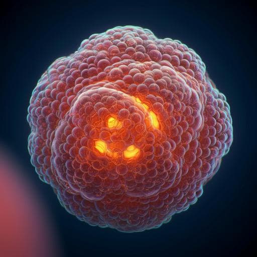
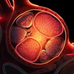
Reconstructing aspects of human embryogenesis with pluripotent stem cells
B. Sozen, V. Jorgensen, et al.
This groundbreaking study, led by Berna Sozen and colleagues, explores early human embryo development using expanded pluripotent stem cells. By creating self-organizing cystic structures that resemble natural embryonic development, the research uncovers the intricacies of blastocyst-like morphology and cell lineage, providing valuable insights into human embryogenesis and its divergence from natural processes.
Related Publications
Explore these studies to deepen your understanding
Adjacent work that informs or extends this paper's methodology and findings.
Medicine and Health
Generation and characterization of cardiac valve endothelial-like cells from human pluripotent stem cells
L. Cheng, M. Xie, et al.
Engineering and Technology
Application of 3D-printed tissue-engineered skin substitute using innovative biomaterial loaded with human adipose-derived stem cells in wound healing
H. Fu, D. Zhang, et al.

Medicine and Health
Critical roles of cytokine storm and bacterial infection in patients with COVID-19: therapeutic potential of mesenchymal stem cells
B. Arjmand, S. Alavi-moghadam, et al.

Medicine and Health
Neuroprotective Effects of Human-Induced Pluripotent Stem Cell-Derived Mesenchymal Stem Cell Extracellular Vesicles in Ischemic Stroke Models
G. Lu, X. Su, et al.


