
Food Science and Technologynpj Science of Food
Obesity, but not high-fat diet, is associated with bone loss that is reversed via CD4+CD25+Foxp3+ Tregs-mediated gut microbiome of non-obese mice
W. Song, Q. Sheng, et al.
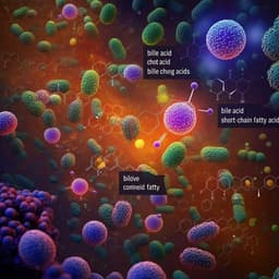
This fascinating study conducted by Wei Song and colleagues reveals the critical link between high-fat diet-induced obesity and bone loss in mice, while highlighting the protective role of a healthy gut microbiome that supports bone health through short-chain fatty acid production. A remarkable fecal microbiota transplantation reversed bone loss, showcasing the gut's power in maintaining skeletal integrity.
Related Publications
Explore these studies to deepen your understanding
Adjacent work that informs or extends this paper's methodology and findings.
Medicine and Health
Distinct signatures of gut microbiome and metabolites associated with significant fibrosis in non-obese NAFLD
G. Lee, H. J. You, et al.

Medicine and Health
Alternate-day fasting delays pubertal development in normal-weight mice but prevents high-fat diet-induced obesity and precocious puberty
R. Ullah, C. Xue, et al.

Medicine and Health
Geraniol reverses obesity by improving conversion of WAT to BAT in high fat diet induced obese rats by inhibiting HMGCoA reductase
S. Chand, A. S. Tripathi, et al.

Food Science and Technology
Short-term *Cudrania tricuspidata* fruit vinegar administration attenuates obesity in high-fat diet-fed mice by improving fat accumulation and metabolic parameters
J. Choi, M. Kim, et al.


