Medicine and HealthNature Communications
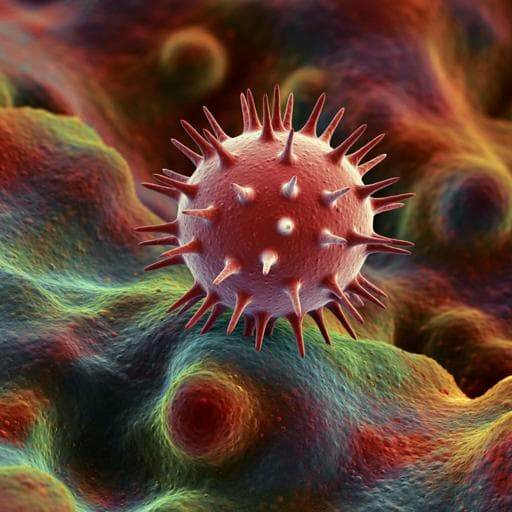
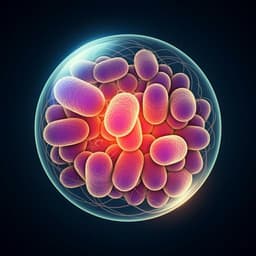
Nanoparticles exhibiting virus-mimic surface topology for enhanced oral delivery
Z. Sang, L. Xu, et al.
Discover the groundbreaking research conducted by Zhentao Sang and colleagues on oral delivery of nano-drug delivery systems! Their innovative core-shell mesoporous silica nanoparticles with virus-like nanospikes demonstrate remarkable improvements in drug delivery, stability, and absorption compared to traditional methods. This study paves the way for enhanced oral therapeutic applications.
Related Publications
Explore these studies to deepen your understanding
Adjacent work that informs or extends this paper's methodology and findings.
Medicine and Health
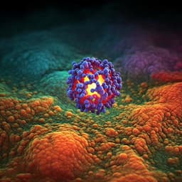
Ligand-switchable nanoparticles resembling viral surface for sequential drug delivery and improved oral insulin therapy
T. Yang, A. Wang, et al.
Engineering and Technology
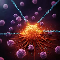
Nanoparticles and convergence of artificial intelligence for targeted drug delivery for cancer therapy: Current progress and challenges
R. P. Singh, A. Natarajan, et al.
Medicine and Health
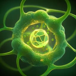
Enhanced plant-derived vesicles for nucleotide delivery for cancer therapy
S. Corvigno, Y. Liu, et al.
Medicine and Health
Programmable probiotics modulate inflammation and gut microbiota for inflammatory bowel disease treatment after effective oral delivery
J. Zhou, M. Li, et al.


