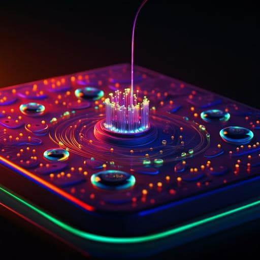
Multiplexed fluorescence and scatter detection with single cell resolution using on-chip fiber optics for droplet microfluidic applications
P. Gupta, A. Mohan, et al.
Explore these studies to deepen your understanding
Adjacent work that informs or extends this paper's methodology and findings.

CAR T-Cell Therapy for Cancer: Latest Updates and Challenges, with a Focus on B-Lymphoid Malignancies and Selected Solid Tumours
H. K. Tang, C. Tang, et al.

Systematic characterization of cleanroom-free fabricated macrovalves, demonstrating pumps and mixers for automated fluid handling tuned for organ-on-chip applications
E. G. B. M. Bossink, A. R. Vollertsen, et al.

On-screen fingerprint sensor with optically and electrically tailored transparent electrode patterns for use on high-resolution mobile displays
H. Kim-lee, S. W. Hong, et al.
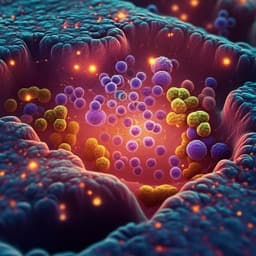
High resolution mapping of the tumor microenvironment using integrated single-cell, spatial and in situ analysis
A. Janesick, R. Shelansky, et al.


