
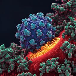
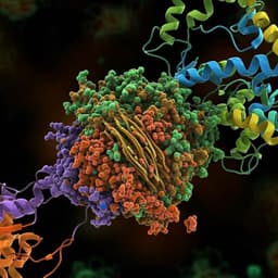
Molecular interaction and inhibition of SARS-CoV-2 binding to the ACE2 receptor
J. Yang, S. J. L. Petitjean, et al.
Explore these studies to deepen your understanding
Adjacent work that informs or extends this paper's methodology and findings.
Broad host range of SARS-CoV-2 and the molecular basis for SARS-CoV-2 binding to cat ACE2
L. Wu, Q. Chen, et al.

Risk factors for and pregnancy outcomes after SARS-CoV-2 in pregnancy according to disease severity: A nationwide cohort study with validation of the SARS-CoV-2 diagnosis of Nordic Federation of Societies of Obstetrics and Gynecology (NFOG)
A. J. M. Aabakke, T. G. Petersen, et al.
Molecular insights into receptor binding energetics and neutralization of SARS-CoV-2 variants
M. Koehler, A. Ray, et al.

Human antibodies to SARS-CoV-2 with a recurring YYDRXG motif retain binding and neutralization to variants of concern including Omicron
H. Liu, C. I. Kaku, et al.


