BiologyLight: Science & Applications
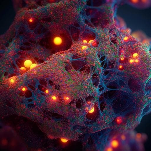
Mid-infrared chemical imaging of intracellular tau fibrils using fluorescence-guided computational photothermal microscopy
J. Zhao, L. Jiang, et al.
Discover the groundbreaking Fluorescence-guided Bond-Selective Intensity Diffraction Tomography (FBS-IDT), developed by Jian Zhao and colleagues, which enables detailed chemical-specific imaging of tau fibrils in their native cellular environment, revealing intriguing links between lipid accumulation and tau aggregation.
Related Publications
Explore these studies to deepen your understanding
Adjacent work that informs or extends this paper's methodology and findings.

Medicine and Health
Specific intracellular signature of SARS-CoV-2 infection using confocal Raman microscopy
H. Salehi, A. Ramoji, et al.

Physics
Three-dimensional imaging of ferroaxial domains using circularly polarized second harmonic generation microscopy
H. Yokota, T. Hayashida, et al.

Biology
Could tau-PET imaging contribute to a better understanding of the different patterns of clinical progression in Alzheimer's disease? A 2-year longitudinal study
J. Lagarde, P. Olivieri, et al.
Biology
Multielement Z-tag imaging by X-ray fluorescence microscopy for next-generation multiplex imaging
M. Strotton, T. Hosogane, et al.


