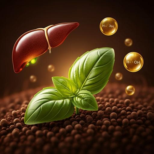
High-fiber basil seed flour reduces insulin resistance and hepatic steatosis in high-fat diet mice
C. Farías, C. Cisternas, et al.
Explore these studies to deepen your understanding
Adjacent work that informs or extends this paper's methodology and findings.

Peripancreatic adipose tissue protects against high-fat-diet-induced hepatic steatosis and insulin resistance in mice
B. Chanclón, Y. Wu, et al.
Maternal high-fat diet programs white and brown adipose tissue lipidome and transcriptome in offspring in a sex- and tissue-dependent manner in mice
C. Savva, L. A. Helguero, et al.

MyD88 determines the protective effects of fish oil and perilla oil against metabolic disorders and inflammation in adipose tissue from mice fed a high-fat diet
F. Wang, M. Hu, et al.

Short-term *Cudrania tricuspidata* fruit vinegar administration attenuates obesity in high-fat diet-fed mice by improving fat accumulation and metabolic parameters
J. Choi, M. Kim, et al.


