Medicine and HealthInternational Journal of Oral Science
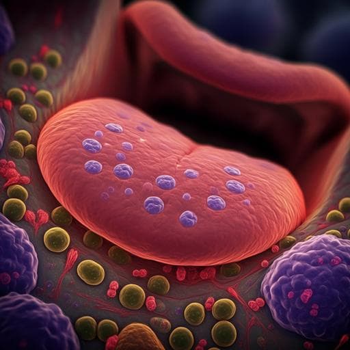
High expression of ACE2 receptor of 2019-nCoV on the epithelial cells of oral mucosa
H. Xu, L. Zhong, et al.
This groundbreaking study explores the role of oral mucosa in the 2019-nCoV infection pathway, highlighting the significant expression of ACE2 in tongue epithelial cells. Conducted by esteemed researchers Hao Xu, Liang Zhong, Jiaxion Deng, Jiakuan Peng, Hongxia Dan, Xin Zeng, Taiwen Li, and Qianming Chen, the findings suggest that the oral cavity may be a critical area for infection risk.
Related Publications
Explore these studies to deepen your understanding
Adjacent work that informs or extends this paper's methodology and findings.

Transportation
Research on the spatial spillover effect of high-speed railway on the income of urban residents in China
Y. Liu, D. Tang, et al.

Business
The impact of risks on small and medium enterprises in the region of Sebha- Libya 2019-2010
M. E. Elgafari and E. A. K. M. Algohaimi

Medicine and Health
Influence of stress induced by the first announced state of emergency due to coronavirus disease 2019 on outpatient blood pressure management in Japan
K. Kobayashi, K. Chin, et al.
Medicine and Health
Effects of maternal diet-induced obesity on metabolic disorders and age-associated miRNA expression in the liver of male mouse offspring
L. V. Mennitti, A. A. M. Carpenter, et al.


