Food Science and TechnologyNature Communications
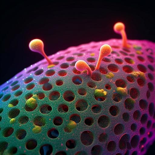
Generation of three-dimensional meat-like tissue from stable pig epiblast stem cells
G. Zhu, D. Gao, et al.
Discover a groundbreaking approach to creating 3D meat-like tissue from porcine pre-gastrulation epiblast stem cells. This innovative study conducted by Gaoxiang Zhu and colleagues showcases a serum-free myogenic differentiation system, paving the way for new cultured meat production techniques.
Related Publications
Explore these studies to deepen your understanding
Adjacent work that informs or extends this paper's methodology and findings.
Medicine and Health
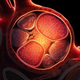
Generation and characterization of cardiac valve endothelial-like cells from human pluripotent stem cells
L. Cheng, M. Xie, et al.

Physics
Three-dimensional imaging of ferroaxial domains using circularly polarized second harmonic generation microscopy
H. Yokota, T. Hayashida, et al.

Medicine and Health
Rapid species identification of pathogenic bacteria from a minute quantity exploiting three-dimensional quantitative phase imaging and artificial neural network
G. Kim, D. Ahn, et al.

Physics
Spin-wave-driven tornado-like dynamics of three-dimensional topological magnetic textures
L. Qiu, L. Shen, et al.


