Medicine and HealthCommunications Biology
Generation and characterization of cardiac valve endothelial-like cells from human pluripotent stem cells
L. Cheng, M. Xie, et al.
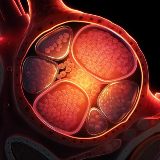
This groundbreaking study introduces a two-step, xeno-free method for generating valve endothelial-like cells from human pluripotent stem cells. The research, conducted by a team including LinXi Cheng and MingHui Xie, reveals the pivotal roles of TGFβ1 and BMP4 in cell differentiation, leading to superior performance on decellularized porcine aortic valve scaffolds. Discover how this advancement could reshape tissue engineering for heart valves!
Related Publications
Explore these studies to deepen your understanding
Adjacent work that informs or extends this paper's methodology and findings.
Food Science and Technology
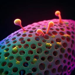
Generation of three-dimensional meat-like tissue from stable pig epiblast stem cells
G. Zhu, D. Gao, et al.

Biology
Reconstructing aspects of human embryogenesis with pluripotent stem cells
B. Sozen, V. Jorgensen, et al.
Medicine and Health
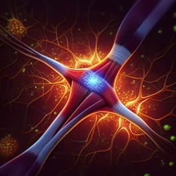
Human sensorimotor organoids derived from healthy and amyotrophic lateral sclerosis stem cells form neuromuscular junctions
J. D. Pereira, D. M. Dubreuil, et al.
Biology
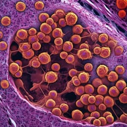
Multiplexed histology analyses for the phenotypic and spatial characterization of human innate lymphoid cells
A. Pascual-reguant, R. Köhler, et al.


