
Veterinary Science
Evaluation of enzymatic protocols to optimize efficiency of bovine adipose tissue-derived mesenchymal stromal cell isolation
E. Heyman, B. Devriendt, et al.
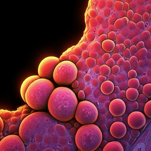
This groundbreaking study by Emma Heyman and colleagues explores innovative isolation techniques for bovine mesenchymal stromal cells from adipose tissue. The team identified 0.1% Liberase™ for 3 hours as the optimal condition, significantly enhancing cell yield and myogenic differentiation, pivotal for cultured meat production.
~3 min • Beginner • English
Introduction
Ensuring sustainable, high-quality protein for a growing global population necessitates alternatives to conventional meat production due to environmental pressures. Cultured meat is a promising solution but faces challenges including limited in vitro proliferation of satellite cells, dependence on animal serum or costly growth factors, and difficulty achieving mature, structured muscle fibers. Mesenchymal stromal cells (MSCs) are explored as alternative sources because they are abundant in adipose tissue (AT), can differentiate into myocytes and adipocytes, and maintain proliferation without losing differentiation potential. To support scalable cultured meat production, maximizing cell yield per tissue isolation is critical. Despite numerous enzymatic protocols to isolate MSCs from AT, comparative data on cell yield are lacking. This study systematically evaluates 32 enzymatic isolation conditions (enzyme type, concentration, incubation time) for bovine subcutaneous AT to identify a protocol that maximizes MSC yield while preserving cell identity and performance, and assesses their myogenic differentiation potential relevant to cultured meat applications.
Literature Review
A focused literature search identified commonly used enzymes and parameters for MSC isolation from AT across species. Collagenase type I is most frequently used; reported enzyme concentrations often center around ~1 mg/mL (≈0.1%), and incubation times around 3 h are common. Other enzymes include trypsin, collagenase types II and IV, and Liberase™. Some studies combined collagenase I with trypsin to isolate ovine or bovine MSCs from abdominal AT. Prior comparative work (e.g., Salehinejad et al., 2012) in human Wharton’s jelly suggested a collagenase/trypsin combination can increase yield and expression of stemness markers compared to other methods. However, across species and sources, isolation efficiency, viability, proliferative capacity, differentiation potential, and marker expression can be affected by enzyme choice, purity, combinations, and process parameters, motivating a systematic, quantitative comparison in bovine AT.
Methodology
Design: Thirty-two isolation conditions were evaluated varying enzyme (Collagenase IA, Collagenase IA + Trypsin 0.25%, Liberase™, Collagenase IV), concentration (0.1% or 0.04%), and incubation time (3 h, 6 h, overnight ≈15 h, 24 h). Eight healthy adult female Belgian White Blue cows (2–4 years) provided subcutaneous AT (back region near shoulder).
Sample handling and digestion: AT was transported (1–2 h) in PBS without Ca2+/Mg2+ with gentamycin, washed, minced, weighed, and digested in 4-well plates at 38.5 °C, 5% CO2 with one of the enzyme conditions. Digestion was neutralized with equal volume cold culture medium (DMEM-LG + 30% FBS, 10−6 M dexamethasone, antibiotics, L-glutamine). After 1 min, the non-buoyant fraction was filtered (70 µm), centrifuged (400 × g, 5 min, RT), washed twice, and resuspended in culture medium. Cells were seeded in T25 flasks, incubated at 38.5 °C, 5% CO2; non-adherent cells were removed after 24 h. Media was changed 2–3×/week. Cells were passaged when ≥5 CFU were observed using trypsin-EDTA.
Success criteria and selection for characterization: An isolation was considered successful if (i) adherent cells at P0 were observed within 21 days, (ii) yield ≥1×10^6 cells/g AT at P1, and (iii) viability >95%. Conditions advancing to further characterization additionally required isolation from ≥5/8 donors and average yield ≥35×10^6 cells/g AT at P1.
Proliferation: From P1 onwards, cells were cultured in expansion medium (culture medium without dexamethasone). During eight passages, cells were seeded at 5,000 cells/cm^2; population doublings (PD) were calculated as ln(Nf/Ni)/ln2, and population doubling time (PDT) as (culture time/PD)×24.
Tri-lineage differentiation: At P4, adipogenic, osteogenic, and chondrogenic differentiation were induced following ISCT-aligned protocols. Adipogenesis used cyclic induction/maintenance media for 14 days and Oil Red O staining; osteogenesis used osteogenic medium for 21 days and Alizarin Red S; chondrogenesis used micromass pellets cultured 21 days in chondrogenic medium with TGF-β3 and Alcian blue staining. Differentiation was quantified via image-based color deconvolution to compute differentiation ratios (differentiation signal area%/nuclear or culture area%).
Immunophenotyping: At P4, multi-color flow cytometry profiled markers including CD29, CD44, CD14, CD34, CD45, CD73, CD79a, MHC II, and CD90 using two panels with appropriate primary/secondary antibodies and controls. At least 10,000 viable singlets were analyzed per sample.
Myogenic differentiation: Cells isolated with the top-yield protocol were differentiated for 14 days in DMEM-HG with 10% FBS, antibiotics, 10 µM 5-aza-2'-deoxycytidine, and 100 nM galectin-1. RT-qPCR assessed PAX3, MYF5, MYOD1, MYF6, MYOG using HPRT1 and YWHAZ as reference genes (validated by NormFinder). Immunofluorescence detected DES, TM, and MyHC.
Statistics: Effects of enzyme concentration and incubation time on yield within enzyme were tested by Kruskal–Wallis with Dunn’s correction. Enzyme effects (fixed at 0.1% and 3 h) were compared to Coll IA by Kruskal–Wallis with Dunn’s post hoc and Bonferroni correction. A Cox proportional hazards frailty model and Kaplan–Meier curves analyzed time to reach sufficient growth (>5 CFU). PDT and flow cytometry data were analyzed by Kruskal–Wallis with Dunn’s post hoc; RT-qPCR and IF by Wilcoxon matched-pairs signed-rank test. Repeated measures ANOVA assessed tri-lineage differentiation versus control. Visualizations were produced in GraphPad Prism.
Key Findings
- All enzymatic conditions yielded spindle-shaped adherent cells within 2 days except Coll IV at 0.04% for 6 h, overnight, and 24 h, which produced no adherent cells even after 21 days. Viability exceeded 95% for all successful conditions.
- Average cell yield across conditions ranged from 4.38×10^6 to 130×10^6 cells/g AT at P1.
- Within each enzyme, varying concentration (0.04% vs 0.1%) or incubation time (3 h, 6 h, overnight, 24 h) did not cause statistically significant differences in yield, though higher yields tended to occur with 0.1% and shorter times (3–6 h).
- Comparing enzymes at 0.1% for 3 h, Liberase™ (0.1%, 3 h) produced significantly higher yields than Collagenase IA (0.1%, 3 h) (p=0.0397). Liberase™ yields ranged 30.48×10^6 to 67.1×10^6 cells/g AT.
- Conditions meeting selection criteria for further characterization: Liberase™ 0.1% (3 h, 6 h, overnight), Coll IA 0.04% (6 h), and Coll IA 0.1% (6 h).
- Time-to-event analysis: >5 CFU was achieved by day 5 for Liberase™ 0.1% (3 h and 6 h) and Coll IA 0.04% (6 h); at day 6 for Liberase™ 0.1% (overnight); and day 7 for Coll IA 0.1% (6 h). Only Liberase™ 0.1% (3 h) produced cells from all donors; other selected conditions failed to yield passagable cells within 21 days for 1–2 donors.
- Proliferation: Cells isolated with Liberase™ 0.1% (3 h and 6 h) showed the lowest PDTs and maintained stable PDT across PD levels, whereas other conditions showed increasing PDT with passaging, indicating better preserved proliferative capacity with Liberase™.
- Tri-lineage differentiation: All selected-condition isolates successfully differentiated into adipocytes, osteocytes, and chondrocytes; no significant inter-condition differences in differentiation ratios.
- Immunophenotype: Bovine AT-MSCs expressed CD29 and CD44 and lacked CD14, CD45, CD73, CD79a, and MHC II across selected conditions, with no significant inter-condition differences.
- Myogenic differentiation (Liberase™ 0.1%, 3 h isolates): After 14 days with 5-aza-2'-deoxycytidine and galectin-1, cells exhibited morphological myogenic features, decreased PAX3 mRNA, and increased MYF5, MYOD1, MYF6, and MYOG mRNA versus undifferentiated controls (p=0.0078). DES, TM, and MyHC proteins were detected in differentiated cells; MyHC was also observed in undifferentiated cells at day 14.
Discussion
Systematic comparison of enzymes, concentrations, and incubation times for bovine subcutaneous AT demonstrated that Liberase™ at 0.1% for 3 h uniquely ensured successful MSC isolation from every donor while delivering high yields and favorable proliferation kinetics. While many enzyme conditions can produce viable MSCs, Liberase™—a blend of purified collagenases I/II and thermolysin—likely digests bovine AT extracellular matrix more efficiently than single collagenases, consistent with the ECM composition of bovine subcutaneous AT (rich in collagens I, III, V, VI; limited collagen IV). Collagenase IV performed poorly under certain conditions, aligning with its lower substrate abundance in this tissue. Within-enzyme parameter tuning (0.04–0.1%; 3 h–24 h) did not significantly alter yield overall, though shorter incubations at 0.1% tended to perform better and prolonged digestion with trypsin combinations reduced yields, consistent with potential over-digestion or membrane protein damage. Importantly, MSC identity and functionality (tri-lineage differentiation, immunophenotypic profile) were preserved across selected conditions. For cultured meat, scaling requires 30–50 population doublings; liberase-isolated cells maintained low PDTs across eight passages, indicating preserved proliferative capacity toward this goal. Myogenic differentiation markers and structural proteins increased under induction, though resultant myotubes remained immature, indicating further bioengineering is needed to achieve mature, well-structured myotubes with robust fusion and contractile protein assembly. Overall, adopting Liberase™ 0.1% for 3 h can improve MSC yield and standardize bovine AT-MSC isolation for downstream applications in cultivated meat.
Conclusion
Liberase™ at 0.1% for 3 h is the most effective enzymatic protocol identified for isolating bovine AT-derived MSCs, ensuring successful recovery from all donors with high yields and preserved proliferative capacity. Isolated cells retained tri-lineage differentiation potential and appropriate MSC immunophenotype, and demonstrated induction of myogenic regulators and structural proteins under myogenic conditions. These findings support bovine AT-MSCs as an attractive cell source for cultured meat production and provide a standardized, efficient isolation protocol. Future work should optimize myogenic differentiation to yield mature, well-structured myotubes with enhanced fusion and contractile function, and evaluate scalability and serum-free/animal component-free conditions.
Limitations
- Donor set was limited to eight healthy adult female Belgian White Blue cows and one anatomical source (subcutaneous AT near the shoulder), which may limit generalizability across breeds, sexes, ages, and adipose depots.
- Only four enzyme conditions (Coll IA, Coll IA + trypsin, Liberase™, Coll IV) with two concentrations and four incubation times were tested; other enzymes, combinations, or mechanical/enzymatic hybrids were not evaluated.
- Some data were summarized without full disclosure (e.g., exact maximum yield 130×10^6 cells/g AT per condition, senescence observations beyond P8 noted as data not shown).
- Myogenic differentiation produced immature myotubes with limited fusion; functional maturation and contractility were not assessed and remain to be optimized.
- All assessments were in vitro; implications for large-scale bioprocessing and serum-free or defined media conditions were not directly tested.
Related Publications
Explore these studies to deepen your understanding of the subject.