Medicine and HealthCell Death and Disease
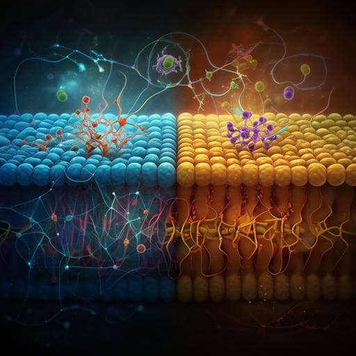
Dual role of Ca²⁺-activated Cl⁻ channel transmembrane member 16A in lipopolysaccharide-induced intestinal epithelial barrier dysfunction in vitro
J. Sui, C. Zhang, et al.
This study unveils the intriguing dual role of TMEM16A in intestinal epithelial barrier function amidst LPS-induced damage. Conducted by a team of researchers from Dalian Medical University, the findings reveal how TMEM16A exacerbates dysfunction with low LPS doses while providing protection at higher doses, challenging our understanding of epithelial responses to inflammation.
Related Publications
Explore these studies to deepen your understanding
Adjacent work that informs or extends this paper's methodology and findings.
Veterinary Science
Dietary prebiotics promote intestinal *Prevotella* in association with a low-responding phenotype in a murine oxazolone-induced model of atopic dermatitis
A. Laigaard, L. Krych, et al.

Medicine and Health
Gliovascular transcriptional perturbations in Alzheimer's disease reveal molecular mechanisms of blood brain barrier dysfunction
Ö. İş, X. Wang, et al.
Engineering and Technology
Towards Novel Biomimetic In Vitro Models of the Blood-Brain Barrier for Drug Permeability Evaluation
I. Mármol, S. Abizanda-campo, et al.

Social Work
Inheritance-induced familial disputes in north-west Ethiopia: the role of legal-policy gaps and aggravating socio-economic dynamics
W. T. Tedla and K. D. Mekonen


