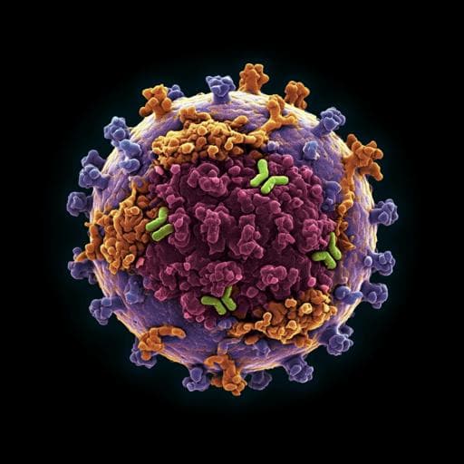
Introduction
Canine parvovirus (CPV), a highly contagious virus that emerged in the mid-1970s, causes severe disease in dogs. It arose from a cross-species transmission event from a feline panleukopenia virus (FPV) ancestor, undergoing mutations that enabled it to infect canine hosts. Subsequent mutations led to the emergence of multiple CPV variants, contributing to its widespread dissemination. The host range of CPV and FPV is largely determined by the virus capsid's ability to bind to the canine transferrin receptor type-1 (TfR). Mutations within the receptor binding site impact both the virus's ability to infect different hosts and its susceptibility to antibody neutralization, highlighting the intricate interplay between receptor binding and antigenic epitopes.
CPV possesses a robust, 26-nm diameter, T=1 icosahedral capsid. Its capsid shell primarily consists of VP2 (~90%) and VP1 (~10%) proteins, created through differential mRNA splicing. The capsid's topology, notably the threefold spike surrounding each icosahedral threefold axis, constitutes a key antigenic site. CPV is a potent antigen, inducing high antibody levels following infection or vaccination. These antibodies effectively neutralize CPV and cross-neutralize related viruses such as FPV and mink enteritis virus, offering protection against infection.
Previous studies have employed various approaches, primarily using panels of rodent monoclonal antibodies (mAbs), to investigate CPV epitopes. These studies identified two major binding regions, A and B, based on antibody competition and the impact of capsid surface mutations on antibody binding. Cryo-EM analyses of Fab-capsid complexes revealed multiple Fab footprints overlapping within these regions, collectively covering ~70% of the capsid's exposed surface. Higher-resolution structures of some of these complexes have been determined, revealing atomic-level interactions. However, these studies primarily used rodent mAbs and didn't fully reflect the canine antibody response to prolonged antigen exposure following infection or vaccination. This study uses advanced cryo-EM techniques to investigate the polyclonal antibody response in dogs after CPV vaccination.
Literature Review
Numerous studies have characterized the antigenic sites of canine and feline parvoviruses using a variety of methods. Rodent monoclonal antibodies (mAbs) have been instrumental in mapping the epitopes, revealing two major binding regions, A and B, on the capsid surface. Cryo-electron microscopy (cryo-EM) studies of Fab-virus complexes have shown that multiple antibodies bind to these regions, with significant overlap between antibody binding sites and the transferrin receptor binding site. These studies primarily utilized rodent mAbs which may not fully reflect the polyclonal antibody response in the natural host. This research advances prior work by employing cryo-EM to analyze the polyclonal antibody response to CPV vaccination in dogs, allowing for a more accurate representation of the natural immune response.
Methodology
The researchers collected blood samples from two beagle puppies at 8 and 12 weeks post-vaccination with a modified live CPV vaccine. Two methods were employed to isolate and prepare the polyclonal Fab fragments for cryo-EM analysis. The first method involved affinity purification of CPV-specific antibodies using a CPV capsid-affinity column, followed by papain digestion to obtain Fab fragments. The second method used protein A/G to isolate total IgGs, followed by papain digestion and purification of the total Fab repertoire using mixed-mode chromatography. In both approaches, the purified Fabs were incubated with CPV capsids to form complexes.
Cryo-EM data were collected for both the affinity-purified and total Fab-virus complexes. Initial icosahedral averaging revealed limited Fab density. To overcome this, the researchers used a custom software, Icosahedral Subparticle Extraction and Correlated Classification (ISECC), to perform subparticle analysis. Subparticles encompassing the A and B sites were extracted and classified, allowing for the identification of distinct Fab populations with different binding orientations. The resulting subparticle maps were refined to near-atomic resolution, providing detailed information on Fab-virus interactions. The capsid and Fab models were built using existing crystal structures as templates, with alanine substitutions used for Fab variable domains due to unknown sequences. Footprints of each Fab on the capsid surface were then mapped.
Key Findings
The cryo-EM analysis revealed a remarkably focused polyclonal antibody response. In both datasets (affinity-purified and total Fab), only one or two distinct Fab populations were identified for each of the A and B epitopes. A total of five unique Fabs were identified across both dogs. The Fab footprints for both A and B site antibodies overlapped with the transferrin receptor (TfR) binding site. This overlap suggests that all identified antibodies could neutralize CPV by sterically hindering TfR binding. The A site antibodies from the two dogs showed some similarities but also differences in their binding angles and footprints on the virus capsid, whereas the B site antibodies showed remarkable similarity, with antibodies from separate dogs exhibiting nearly identical binding orientations but with flipped heavy and light chain arrangements. The study also noted steric clashes between some of the antibodies bound to the capsid, limiting the number of antibodies bound per capsid. Affinity purification was found not to be strictly necessary, as results using total Fab were consistent with those using affinity-purified Fab.
Discussion
This study demonstrates a novel approach for mapping polyclonal antibody responses to icosahedral viruses using cryo-EM and subparticle analysis. The results reveal a surprisingly limited yet potentially redundant polyclonal response to CPV vaccination. While only a small number of distinct antibodies were identified, their binding sites overlap significantly with the TfR binding site, suggesting a robust neutralizing effect. This approach avoids the limitations of traditional methods using excess monoclonal antibodies, offering a more realistic representation of the in vivo immune response. The focused nature of the response might be influenced by the capsid topology, the exposed nature of the B site, or specific features of canine B-cell recognition. Further studies are needed to fully understand the factors contributing to this focused response. The methodology presented here is applicable to studying polyclonal antibody binding to other icosahedral viruses.
Conclusion
This study provides a high-resolution structural view of the polyclonal antibody response to CPV vaccination in dogs. The findings highlight a surprisingly limited yet effective neutralizing antibody response, with a small number of distinct antibodies targeting key epitopes that overlap with the TfR binding site. The developed methodology, utilizing cryo-EM and subparticle analysis, offers a powerful tool for studying polyclonal antibody binding to icosahedral viruses. Future research could explore the broader implications of these findings for vaccine design and the understanding of virus-host interactions in other systems.
Limitations
The study used a limited sample size (two dogs) and focused on a specific time point post-vaccination. Therefore, the findings might not be fully generalizable to all canine populations or different time points during the immune response. The use of alanine substitutions in the Fab variable domains, due to the unknown antibody sequences, might have resulted in an underestimation of actual antibody-virus contacts. The reliance on a modified live vaccine might also limit the generalizability of the results to responses elicited by other vaccine types or by natural infection.
Related Publications
Explore these studies to deepen your understanding of the subject.