
Medicine and Health
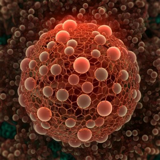
An instantly fixable and self-adaptive scaffold for skull regeneration by autologous stem cell recruitment and angiogenesis
G. Lu, Y. Xu, et al.
Discover an innovative scaffold that addresses cranial defect reconstruction challenges! Developed by Gonggong Lu and colleagues, this self-adaptive scaffold enhances bone regeneration, promotes macrophage polarization, and integrates seamlessly with the biological environment, achieving impressive bone cover in model tests.
~3 min • Beginner • English
Introduction
The study addresses the challenge of reconstructing large craniofacial bone defects where current solutions (autografts/allografts, titanium mesh, PEEK) face limitations including donor site morbidity, poor stretchability, weak osteointegration, high stiffness, and surgical complexity. Craniofacial bones form via intramembranous ossification and require coordinated osteogenesis and angiogenesis, yet many regeneration strategies fail to yield highly vascularized bone due to instability, poor wet adhesion, and limited functional cell settlement. Hydrogel-based adhesives and growth factor or cell delivery approaches have barriers including poor wet adhesion, limited stem cell sources, cost, and regulatory hurdles. The authors propose a cell/factor-free, instantly fixable scaffold that mimics the native bone niche to recruit endogenous stem cells (ESCs), modulate immunity, and initiate angiogenesis and osteogenesis immediately after craniectomy. Inspired by mussel adhesion, they integrate dopamine-modified hyaluronic acid to chelate calcium from micro-hydroxyapatite and covalently link type I collagen, forming a hybrid cross-linked scaffold that is flexible, self-adhesive in wet environments, and mechanically matched to skull tissue.
Literature Review
Background literature highlights: (1) Autologous/allogeneic grafts are limited by donor morbidity and cost; synthetic substitutes (titanium, PEEK) often have high modulus, poor osteointegration, and frictional issues. (2) Intramembranous ossification depends on mesenchymal cell differentiation with tight coupling to angiogenesis; existing scaffolds often fail to produce vascularized bone. (3) Hydrogel adhesives can improve tissue adhesion and cell retention, but commonly lack strong wet adhesion and efficient cell recruitment. (4) Pro-angiogenic strategies using VEGF delivery or exogenous cells face availability, cost, translation, and regulatory constraints. (5) Mussel-inspired catechol chemistry offers strong wet adhesion and metal chelation. Prior studies show immunomodulation (M2 polarization), macrophage-derived VEGF/BMP-2 roles, and the impact of scaffold microstructure on ESC recruitment and osteogenesis/angiogenesis. These inform the design of a hybrid cross-linked, dopamine-mediated scaffold to emulate bone ECM and mobilize ESCs.
Methodology
Materials and scaffold fabrication: Dopamine-modified hyaluronic acid (HAD) was synthesized by EDC/NHS coupling of dopamine to hyaluronic acid (MW ~0.3 MDa), achieving 7.8% catechol substitution (1H NMR, UV, FTIR). Scaffolds included: HAD; type I collagen (Col I); dual cross-linked scaffold (DCLS: HAD + Col I); and hybrid cross-linked scaffold (HCLS: HAD + Col I + micron hydroxyapatite, µHAp). HCLS was formed by dispersing µHAp (average ~14.45 µm, Ca/P 1.67) in HAD, mixing with Col I under ice, and rapidly adjusting pH to 7.4 to induce gelation and prevent particle settling. DCLS formed similarly without µHAp.
Characterization: µHAp crystallinity (XRD), size and morphology (DLS, SEM/EDS). Chemical structure and bonding assessed by FTIR, UV, 1H NMR, XPS (catechol oxidation, Michael addition with Col I amines; Ca2+ chelation evidenced by Ca2p signal in HCLS and loss of P2p; sustained chelation after 0.3 M CaCl2 incubation). Thermal properties via DSC (denaturation temperature) and TGA (component content; µHAp in HCLS ~19% w/w). Microstructure via SEM; porosity and pore size quantified (HCLS: 87±9% porosity; 359±49 µm pores). Component distribution validated by Masson's trichrome (collagen) and calcein staining (µHAp) using CLSM. Swelling in DPBS (14 days) and enzymatic degradation (hyaluronidase/collagenase). Mechanical testing: tensile and compressive DMA (G') at 5 Hz; tests with and without blood infiltration to mimic wet implantation; adhesion strength on substrates (glass, Ti, skull, pigskin, polyethylene).
In vivo interface mechanics: Rabbit cranial defect model (Ø 9 mm) assessed at days 7 and 14 for interface integration (gross, SEM), mineralization (SEM, EDS Ca/P), push-out tests (interfacial binding force), and implant storage modulus (DMA).
Immunomodulation assays: In vitro RAW 264.7 culture on DCLS vs HCLS; SEM for morphology, IF for M1 (CD197) and M2 (CD206) markers; semiquantitative analysis. In vivo mouse intramuscular implantation (5 mm discs) of DCLS and HCLS; H&E histology, flow cytometry for macrophage recruitment and polarization (F4/80, CD197, CD206), ELISA of cytokines (TNF-α, IL-1β, IL-4, IL-10, IL-1ra), IF for F4/80 co-localized VEGF and BMP-2, and CD197/CD206. Similar analyses in rabbit skull defects at 7 and 14 days.
BMSC osteogenesis and ectopic mineralization: Rabbit BMSCs encapsulated in hydrogels; viability (live/dead), proliferation (CCK-8), morphology (CLSM cytoskeleton; SEM), IHC for BMP-2 and VEGF, qRT-PCR for Runx2, OCN, OPN at days 3, 7, 14. Subcutaneous implantation of BMSC-laden hydrogels in nude mice (4 weeks): gross, DMA, micro-CT 3D reconstructions, quantitative apatite volume (AV) and AV/TV, SEM/EDS (Ca/P), XRD, IF for Runx2 and CD31, IHC for VEGF.
ESC recruitment assays: In vitro adhesion and migration: scaffolds incubated with total rabbit cranial bone marrow cell suspension (48 h), IF for CD44, cytoskeleton, morphology; transwell migration assay with scaffolds in lower chamber to quantify migrated BMSCs (crystal violet OD at 573 nm). In vivo ESC recruitment in rabbit cranial defects (Ø 9 mm): at 1 week, SEM and CLSM for cytoskeleton; IF for CD44, CD90, Runx2, BMP-2; dual turntable confocal quantification of total cells and CD44+ ESCs; CD31 IF for vascular infiltration and density.
Transcriptomics: RNA-seq of BMSCs cultured 14 days in Col I, DCLS, HCLS (n=3/group). Analyses included Pearson correlation, PCA, differential expression (volcano plots), GO and KEGG enrichment, and PPI networks, focusing on cellular activity, osteogenesis, angiogenesis, and ESC recruitment pathways.
In vivo cranial regeneration: Rabbit model (Ø 9 mm) with groups: blank, DCLS, HCLS, normal; endpoints 4 and 12 weeks. Assessments: micro-CT (BV/TV, BMD, trabecular thickness, number, spacing, bone cover area), X-ray, SEM cross-sections, push-out tests, qPCR (Col I, Runx2, BMP-2, OCN, OPN, VEGF), Western blot (Runx2, BMP-2, VEGF), histology (H&E, Masson's trichrome), CD31 IF, IHC (BMP-2, Runx2, OCN, VEGF).
Large animal model: Beagle dog cranial defects (Ø 15 mm), HCLS vs blank; endpoints 4 and 12 weeks. X-ray, micro-CT (BV/TV, new bone volume, bone cover ratio), histology (H&E, MT), and CD31 IF with semiquantitative analysis.
Key Findings
- Chemistry and structure: HCLS formed via dopamine-mediated hybrid cross-linking among HAD, Col I, and µHAp. XPS showed Ca2+ chelation; FTIR indicated consumption of phenolic OH; DSC denaturation temperature highest for HCLS (113.5 °C) vs Col I (84.6 °C), HAD (88.2 °C), DCLS (111.7 °C). HCLS contained ~19% w/w µHAp.
- Architecture and mechanics: HCLS exhibited high porosity (87±9%) and pore size (359±49 µm). Minimal size change in PBS over 14 days (95±8%), versus swelling of HAD (175±10%) and contraction of Col I (26±5%). HCLS storage modulus G' remained stable (∼90–85 kPa) even after blood infiltration; tensile strength at breakdown 34.8 kPa; strong wet adhesion to diverse substrates; instant fixation to wet skull.
- Interface integration: In rabbit defects, HCLS showed tight host interface by day 7 with increasing mineralized nanolath-like apatite by day 14; push-out force increased to 32±3 N at day 14; implant G' increased to 60±3 kPa.
- Immunomodulation: In vitro, HCLS enhanced macrophage spreading, increased CD206 (M2) and reduced CD197 (M1). In mouse intramuscular model, HCLS decreased macrophage infiltration over time, increased M2 (CD197−CD206+) and decreased M1 (CD197+CD206−), downregulated TNF-α and IL-1β, upregulated IL-4, IL-10, IL-1ra. F4/80+ macrophages co-expressed higher VEGF and BMP-2 in HCLS in both mouse and rabbit models.
- BMSC osteogenesis and ectopic mineralization: BMSCs in HCLS showed highest viability and proliferation, enhanced spreading, and strongest BMP-2/VEGF staining. Osteogenic genes (Runx2, OCN, OPN) were significantly upregulated over 14 days. In nude mice, BMSC-laden HCLS increased storage modulus to 103±8 kPa (~5× baseline), with micro-CT showing AV 30±2.3 mm³ and AV/TV 78±4.1%. SEM/EDS revealed embedded ∼200 nm mineral nanoparticles with Ca/P ≈1.91±0.34 and XRD peaks consistent with HAp. Runx2 and CD31 signals were highest in HCLS.
- ESC recruitment: In vitro, HCLS anchored more CD44+ cells and induced greater BMSC migration (transwell OD increases; p<0.01–0.001). In vivo (rabbit, 1 week), HCLS contained 38,900 total cells vs 30,650 (DCLS) and 20,470 (HAD); CD44+ ESC ratio 66.1% (HCLS) vs 34.2% (DCLS) and 31.9% (HAD). Higher CD90+ cell presence confirmed BMSC-like recruitment. Vascular density in HCLS at 1 week reached 1.97±0.54 mm/mm², with more central infiltration.
- Transcriptomics: Relative to Col I, DCLS upregulated genes in ECM organization, chemotaxis, adhesion, angiogenesis, and ossification; KEGG pathways included cytokine/chemokine interactions, focal adhesion, NF-κB and PPAR signaling (M2 polarization), PI3K-Akt, HIF-1, TGF-β. Adding µHAp (HCLS vs DCLS) further enriched PI3K-Akt, focal adhesion, Rap1, cell cycle, and inflammatory regulation (phospholipase D–NF-κB), and upregulated genes associated with ESC recruitment (e.g., CXCL12, LAMA2/4, SRGN), angiogenesis (PGF, IGF1, PDGFRA, ADAMTS3), and osteogenesis (OMD, OGN, GREM1, COL14A1).
- Rabbit cranial regeneration (Ø 9 mm): At 4 weeks, BV/TV: blank 18.7±3.5%, DCLS 23.5±3.7%, HCLS 47.3±4.4%. At 12 weeks, HCLS BV/TV 79.6±7.3% vs DCLS 64.0±5.7% and blank 19.1±2.1%; BMD 238 mg HA/cm³; bone cover area 97%; trabecular thickness 0.28 mm; trabecular number 1.75 mm−1; trabecular spacing reduced to 0.37 mm. Push-out breaking load increased from 36.7 N to 68.5 N (near normal 80.5 N). qPCR and WB showed highest Col I, Runx2, BMP-2, OCN, OPN, and VEGF gene/protein expressions in HCLS (VEGF protein slightly lower than DCLS despite higher gene levels). Histology and CD31 IF confirmed mature lamellar bone and dense neovascularization.
- Beagle dog cranial regeneration (Ø 15 mm): HCLS increased BV/TV to 35.7±5.1% (4 w) and 58.7±7.6% (12 w); new bone volume 126.1±15 mm³ (4 w) and 207.1±27.3 mm³ (12 w); bone cover ratio 71.7±6.9% at 12 w. Histology showed progressive bone and collagen maturation and significantly higher CD31+ neovascularization versus blank.
Discussion
The hybrid cross-linked, dopamine-mediated scaffold (HCLS) effectively addresses the need for an immediately implantable, wet-adhesive, and mechanically compatible cranial implant. By mimicking bone ECM composition (Col I + HAp) and incorporating catechol chemistry, HCLS achieves instant wet adhesion, flexible deformation to accommodate intracranial pressure, and strong host-scaffold interface integration. This stable, porous niche promotes macrophage M2 polarization and secretion of VEGF and BMP-2, which together drive early angiogenesis and osteoinduction. Concurrently, HCLS recruits endogenous stem/progenitor cells (CD44+, CD90+) from surrounding tissues and marrow, supports their adhesion and proliferation, and enhances osteogenic differentiation. Transcriptomic analyses corroborate upregulated pathways in ECM organization, cell adhesion (focal adhesion, integrin binding), PI3K-Akt signaling, and immunomodulatory routes (NF-κB, PPAR), providing mechanistic insight into coupled osteogenesis and angiogenesis. In rabbits, this translates into near-complete bone defect closure with high bone quality metrics by 12 weeks, while in beagle dogs substantial regeneration is achieved though somewhat reduced, likely due to a more demanding mechanical environment. Overall, HCLS demonstrates that a cell- and factor-free strategy can leverage host immunity and endogenous stem cells to regenerate vascularized cranial bone, with potential for single-stage craniectomy and cranioplasty.
Conclusion
An instantly fixable, self-adaptive hybrid cross-linked scaffold (HCLS) composed of dopamine-modified hyaluronic acid, type I collagen, and micro-hydroxyapatite enables robust wet adhesion, mechanical compliance, and interface integration in cranial defects. HCLS modulates macrophage polarization toward M2, recruits endogenous stem cells, and accelerates angiogenesis and osteogenesis, achieving 97% bone coverage in rabbit (Ø 9 mm) and ~72% in beagle dog (Ø 15 mm) models at 12 weeks. The scaffold’s biomimetic architecture and catechol-mediated interactions underpin enhanced cellular activity and pro-regenerative gene expression. Future work should optimize structural and adhesive design to better withstand the dynamic mechanical environment of large animal and human skulls, evaluate long-term stability and remodeling, and advance towards clinical translation as a cell/factor-free, single-stage cranial reconstruction approach.
Limitations
- Large animal performance was weaker than in rabbits, attributed to higher mechanical loads and motion from skull musculature; without exogenous fixation, HCLS adhesion may be insufficient for long-term stability in highly mobile sites. Structural and adhesive optimization is needed.
- DCLS degraded relatively quickly, limiting structural integrity and vascularization, highlighting sensitivity to degradation kinetics.
- Discrepancies between VEGF mRNA and protein levels suggest complex regulation and time/space differences between transcription and translation that warrant further temporal profiling.
- Translation to human skull (thicker, stiffer, different mechanical milieu) remains to be validated; long-term durability and safety beyond 12 weeks were not assessed.
- While CD44/CD90 suggest ESC/BMSC recruitment, marker overlap with other cell types could confound precise lineage attribution without fate mapping.
Related Publications
Explore these studies to deepen your understanding of the subject.