
Medicine and Health
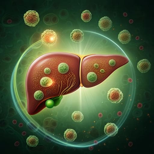
Alantolactone attenuates high-fat diet-induced inflammation and oxidative stress in non-alcoholic fatty liver disease
J. Wang, Y. Jiang, et al.
This study reveals the promising therapeutic effect of Alantolactone (Ala) on Nonalcoholic fatty liver disease (NAFLD). By inhibiting inflammation, fibrosis, and oxidative stress in mice, Ala showcases its potential as a treatment option for NAFLD, suggesting a novel approach to tackling this condition.
~3 min • Beginner • English
Introduction
Obesity-related diseases are rising globally, and NAFLD prevalence continues to increase. NAFLD ranges from simple steatosis to non-alcoholic steatohepatitis (NASH), characterized by inflammation, hepatocellular injury, and fibrosis, and can progress to cirrhosis and hepatocellular carcinoma. No FDA-approved drugs exist for NASH. Chronic inflammation and oxidative stress are central to NAFLD progression. Steatosis activates NF-κB via IKKβ, inducing pro-inflammatory cytokines (TNF-α, IL-6, IL-1β). Excess liver fat causes lipotoxicity, mitochondrial dysfunction, ER stress, and ROS generation, promoting apoptosis, lipid peroxidation, inflammation, and fibrogenesis. Alantolactone (Ala), a natural compound from Inula helenium L., exhibits anti-inflammatory, antitumor, antibacterial, and neuroprotective activities, notably via NF-κB inhibition and modulation of Nrf2/HO-1 in prior models. Whether Ala has therapeutic effects in NAFLD was unknown. This study aimed to test Ala’s hepatoprotective effects in NAFLD and its mechanisms, focusing on inflammation and oxidative stress.
Literature Review
The authors frame NAFLD pathogenesis around intertwined inflammation and oxidative stress, highlighting NF-κB-driven cytokine production and ROS-induced damage as key drivers of progression from steatosis to fibrosis. They note the absence of FDA-approved pharmacotherapies for NASH and discuss natural-product strategies. Prior research shows alantolactone exerts anti-inflammatory effects across models, including diabetic nephropathy (via NF-κB inhibition), cigarette smoke-induced bronchial epithelial injury (via Nrf2/HO-1 activation and NF-κB inhibition), neuroinflammation, and colitis (potentially via PXR and NF-κB suppression). However, its role in liver inflammatory disease/NAFLD had not been explored. The discussion also cites evidence linking Nrf2/Keap1 signaling to NAFLD protection and suggests NOD-like receptor signaling as another potentially relevant pathway modulated by Ala.
Methodology
In vivo model: Male C57BL/6 mice (18–20 g) were housed under standard conditions. Diets: low-fat diet (LFD; 10% kcal fat) or high-fat diet (HFD; 60% kcal fat). Groups (n=6 each): (i) CON: LFD + vehicle (0.5% CMC-Na); (ii) Ala 10 mg/kg: LFD + alantolactone; (iii) HFD + vehicle; (iv) HFD + Ala 5 mg/kg; (v) HFD + Ala 10 mg/kg. HFD or LFD feeding lasted 16 weeks, followed by 8 weeks of Ala or vehicle by oral gavage once every 2 days. Mice were anesthetized with sodium pentobarbital (i.p., 0.2 mL at 100 mg/mL) before tissue/serum collection. Outcomes: Body and liver weight; serum ALT, AST; serum lipids (TG, TCH, LDL-C); histology (H&E, Oil Red O, Sirius Red, Masson’s trichrome); ROS by DHE staining; SOD activity and MDA levels; liver mRNA (RT-qPCR) and protein (Western blot) for fibrosis markers (COL1, TGF-β1, α-SMA) and redox/inflammation pathway proteins (Nrf2, HO-1, Keap1; p-p65, p65, IκBα; nuclear p65). In vitro model: AML-12 hepatocytes cultured in DMEM/F12 with supplements. Cytotoxicity assessed by MTT after 48 h across Ala 0.2–50 µM to select non-toxic doses; 1 and 5 µM were used. Lipotoxicity induced with palmitic acid (PA) 200 µM. Treatments: Ala pretreatment (2 h) prior to PA exposure (1 h for NF-κB activation, 12 h for cytokine mRNA, 24 h for fibrosis markers). Nrf2 inhibitor ML385 (4 µM) was used to probe dependence on Nrf2 signaling. Assays: DHE for ROS; Western blots for Nrf2, HO-1, Keap1; NF-κB pathway proteins and nuclear p65; RT-qPCR for Tnf, Il6, Il1b; Col1a1, Tgfb1, Acta2; secreted IL-6 by ELISA (supplementary). RNA-seq: Liver RNA extracted with TRIzol; poly(A) selection; fragmentation; cDNA synthesis (SuperScript II); sequencing on Illumina NovaSeq 6000. Differential expression called with thresholds fold change >2 or <0.5 and p<0.05. Pathway enrichment via modEnrichr (BioPlanet 2019). Statistics: Data are mean ± SEM. Normality assessed by Shapiro–Wilk. Two-group comparisons by Student’s t-test; multiple groups by one-way ANOVA with Dunnett’s post hoc test. Significance at p<0.05.
Key Findings
- Ala improved liver injury without affecting body weight in HFD-fed mice. Serum ALT and AST were reduced vs HFD controls; liver weight gain was attenuated (n=6/group; significance indicators typically ** or ***). - Lipid metabolism: Ala lowered serum TG, TCH, and LDL-C in HFD mice. Hepatic steatosis was reduced by Ala, evidenced by fewer/lower-volume lipid droplets on H&E and Oil Red O staining, and decreased hepatic expression of lipogenesis genes Fasn, Acaca, and Srebp1. - Fibrosis: Ala decreased collagen deposition in HFD livers (Sirius Red and Masson’s trichrome quantification), and reduced fibrosis markers at protein (COL1, TGF-β1, α-SMA) and mRNA (Col1a1, Tgfb1, Acta2) levels. - Oxidative stress and Nrf2/Keap1: RNA-seq identified 1,446 DEGs between HFD and control (1,074 up, 372 down) and 912 DEGs between HFD+Ala 10 mg/kg and HFD (712 up, 200 down). Forty genes overlapped as Ala-controlled in the HFD context, with pathway analysis (BioPlanet 2019) implicating Keap1–Nrf2 among top hits. In HFD liver, Nrf2 and HO-1 proteins were decreased and Keap1 increased; Ala reversed these. Ala increased mRNA of Nfe2l2, Nqo1, Hmox1. DHE staining showed reduced hepatic ROS with Ala; SOD activity increased and MDA decreased. - Inflammation and NF-κB: HFD increased phosphorylated p65 and decreased IκBα, with enhanced p65 nuclear translocation; Ala suppressed p65 phosphorylation and nuclear localization and preserved IκBα. Proinflammatory cytokine mRNAs (Tnf, Il6, Il1b) and serum IL-6 were reduced by Ala. - In vitro AML-12 findings: Non-toxic Ala doses (1 and 5 µM) selected. PA (200 µM) increased ROS and impaired antioxidant defense (↓Nrf2/HO-1, ↑Keap1); Ala pretreatment reduced ROS and restored Nrf2/HO-1 while lowering Keap1. The Nrf2 inhibitor ML385 (4 µM) exacerbated PA-induced inflammation/fibrosis and partially blunted Ala’s anti-inflammatory and antifibrotic effects, indicating Nrf2 involvement, though Ala retained some protection. Ala inhibited NF-κB activation (↓p-p65, preserved IκBα, ↓nuclear p65) and reduced PA-induced Tnf, Il6, Il1b mRNAs and secreted IL-6. Ala also suppressed PA-induced fibrosis markers at mRNA (Col1a1, Tgfb1, Acta2) and protein (COL1, TGF-β1, α-SMA) levels. Overall, Ala confers hepatoprotection in NAFLD models by reducing steatosis, inflammation, oxidative stress, and fibrosis, associated with activation of Nrf2/Keap1 and inhibition of NF-κB.
Discussion
The study addresses whether alantolactone can therapeutically mitigate NAFLD pathology. Using HFD-fed mice and PA-challenged hepatocytes, Ala consistently reduced liver injury, steatosis, inflammation, oxidative stress, and fibrosis. Mechanistically, Ala activated the Nrf2/Keap1 antioxidant pathway (↑Nrf2, ↑HO-1, ↓Keap1; increased antioxidant gene expression; improved SOD/MDA, reduced ROS) and inhibited NF-κB signaling (reduced p65 phosphorylation and nuclear translocation; restored IκBα), thereby disrupting the deleterious feedback loop between oxidative stress and inflammation. RNA-seq supported pathway-level modulation, highlighting Keap1–Nrf2 involvement among Ala-responsive genes in the HFD context. The partial loss of Ala’s benefits with Nrf2 inhibition (ML385) indicates that Nrf2 activation is a key, but not exclusive, mediator of Ala’s effects. The findings complement prior reports of Ala’s anti-inflammatory actions in other tissues and suggest broader hepatic benefits, potentially including modulation of additional pathways such as NOD-like receptor signaling. Collectively, the data support Ala as a candidate therapeutic for NAFLD by targeting intertwined inflammatory and oxidative mechanisms driving disease progression.
Conclusion
Ala protects against NAFLD in vivo and in vitro by reducing inflammation, oxidative stress, steatosis, and fibrosis. Its benefits are closely linked to suppression of NF-κB-mediated inflammation and activation of the Nrf2/Keap1 antioxidant pathway. These multifaceted effects position alantolactone as a promising therapeutic candidate for NAFLD. Future research should identify direct molecular targets of Ala, validate efficacy in additional fibrosis models (e.g., CCl4), assess metabolic outcomes such as glucose and insulin tolerance and high-sucrose diet models, evaluate effects on adipose tissue, and further interrogate potential roles of NOD-like receptor signaling.
Limitations
- Molecular targets of alantolactone remain undefined; although Ala modulates NF-κB and Nrf2/Keap1, direct binding targets were not determined. - The NF-κB pathway did not emerge in pathway enrichment (BioPlanet), possibly due to limited gene overlap/sample size. - Metabolic phenotyping was incomplete: no glucose or insulin tolerance tests were performed; effects in high-sucrose diet models are hypothesized but untested. - The HFD model, while accepted for NAFLD, has limitations for advanced fibrosis; additional validation in fibrosis-centric models (e.g., CCl4) is warranted. - Adipose tissue was not collected, preventing assessment of Ala’s impact on adipose lipolysis and cross-talk. - In vitro dependence on Nrf2 was probed pharmacologically (ML385) but genetic validation (e.g., Nrf2 knockdown/knockout) was not performed.
Related Publications
Explore these studies to deepen your understanding of the subject.