Medicine and Health
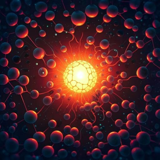
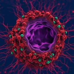
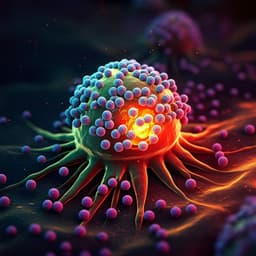
Activatable polymer nanoagonist for second near-infrared photothermal immunotherapy of cancer
Y. Jiang, J. Huang, et al.
Discover the groundbreaking research by Yuyan Jiang, Jiaguo Huang, Cheng Xu, and Kanyi Pu on a novel photothermally activatable polymeric pro-nanoagonist (APNA) that enhances combinational photothermal immunotherapy for cancer. This innovative approach utilizes deep-tissue-penetrating NIR-II light to trigger tumor ablation and boost systemic antitumor immunity.
~3 min • Beginner • English
Introduction
The study addresses the challenge of precisely controlling cancer immune activation in space and time to improve efficacy and safety of immunotherapy. While nanomedicine can enhance delivery and enable combination therapies, many immunotherapeutic nanoagents rely on endogenous biochemical cues (pH, ROS, enzymes), which limits bioavailability and selectivity. External stimuli like near-infrared (NIR) light offer independent, precise control; NIR-II (1000–1300 nm) has superior tissue penetration and lower phototoxicity than NIR-I. The authors hypothesize that a semiconducting polymer nanoagent that converts NIR-II light to heat can both ablate tumors and thermally uncage a covalently linked toll-like receptor 7/8 agonist (R848), achieving spatiotemporally confined immune activation deep within tumors, thereby enhancing systemic antitumor immunity and suppressing metastasis.
Literature Review
- Immunotherapy has transformed cancer treatment but can have limited response rates and immune-related adverse effects.
- Nanomedicine improves immunotherapy via tunable properties, optimized biodistribution/retention, and facile integration with other modalities (chemo-, photo-, radiotherapy).
- Smart immunotherapeutic nanoagents responsive to endogenous biomarkers (pH, ROS, enzymes) have been developed, e.g., disulfide-responsive IL-15 super-agonist complexes; pH-responsive PD-1 Ab conjugates. However, reliance on pathological vs basal biomarker differences limits selectivity and bioavailability.
- External stimuli enable precise spatiotemporal control. NIR-I-responsive systems have triggered inhibitor activation or nucleic acid release. NIR-II offers deeper penetration and lower permissible exposure limits, but few NIR-II nanotransducers exist (gold nanostructures, transition metal nanoparticles, naphthalocyanines).
- Semiconducting polymer nanoparticles (SPNs) have emerged as promising organic NIR-II agents with excellent photothermal performance and biocompatibility, used for deep-tissue photoacoustic imaging, photothermal therapy, and ferrotherapy. Prior to this work, SPN-based NIR-II photoregulation of immunotherapy had not been demonstrated.
Methodology
- Synthesis:
• Semiconducting polymer precursor PBODO-Br synthesized by Stille polycondensation of BBT, DPP-Br, and OT, yielding a NIR-II absorbing backbone. Converted to PBODO-N3 via azide substitution.
• Thermolabile linker VA-044 modified with 7-iodoheptanoic acid and coupled to R848 via Steglich esterification to obtain caged agonist VR. A bifunctional alkyne-PEG-NH2 was coupled to VR (EDC/NHS) to form alkyne-PEG-VR.
• PBODO-N3 clicked with alkyne-PEG-VR to yield pBODO-PEG-VR (pro-agonist polymer). Control pBODO-PEG synthesized similarly with methoxy-PEG-alkyne.
• Self-assembly in aqueous media generated APNA (from pBODO-PEG-VR) and APNC (from pBODO-PEG) nanoparticles. NCBS dye (2.5 wt%) was doped for fluorescence tracking in some experiments.
- Characterization:
• UV–vis–NIR absorption spectra; DLS for hydrodynamic size; TEM for morphology; zeta potential; HPLC for drug loading; photothermal performance at 1064 nm (IR camera) and photostability over heating/cooling cycles; photothermal conversion efficiency calculated from heating/cooling curves (η ≈ 84.4%).
• Photothermal activation: APNA irradiated at 1064 nm; products analyzed by HPLC, LCMS, 1H NMR; subsequent esterase hydrolysis to liberate R848; quantified activation vs temperature (37, 45, 55 °C) and time.
- In vitro studies:
• Cellular uptake: Confocal imaging of 4T1 cancer cells and BMDCs incubated with APNAf/APNCf; lysosomal colocalization assessed with LysoTracker; DAPI nuclear stain.
• Cytotoxicity/PTT: 4T1 cells treated with APNA or APNC (0–50 µg mL⁻1), with/without 1064 nm irradiation (1 W cm⁻2, 6 min); MTS assay at 24 h.
• DC maturation: APNA or APNC solutions pre-irradiated (1064 nm, 10 min) then incubated with immature BMDCs; flow cytometry for CD11c+CD80+CD86+ maturation; free R848 as control. Mixed lymphocyte reaction measured CD3+CD8+ T cells and IFN-γ by ELISA.
- In vivo studies (Balb/c):
• Biodistribution/accumulation: IV injection of APNAF/APNCF; whole-body NIR fluorescence imaging over 32 h; ex vivo organ fluorescence.
• Therapy: Bilateral 4T1 model (primary right flank; distant left flank inoculated 5 days later). IV injection of APNA or APNC (PBODO 250 µg mL⁻1, 200 µL); 24 h later, primary tumor irradiated with 1064 nm laser (1 W cm⁻2, 10 min). Tumor surface temperature monitored; primary and distant tumor volumes tracked for 30 days; survival; body weight.
• Histology: H&E staining of primary tumors; assessment of lung and liver metastases (nodule counts and H&E).
• Immune activation: Flow cytometry of tumor-draining lymph node DC maturation (CD45+CD11c+CD80+CD86+); serum cytokines (IL-6, IL-12p70, IFN-γ, TNF-α) by ELISA at days 1, 3, 7; T cell infiltration (CD8+, CD4+) in primary and distant tumors, spleen, and blood; tumor-specific CTL generation and cytotoxicity assays.
• T-cell dependence: Replicated therapy in T-cell-deficient NCr nude mice.
- Mechanistic study in deep tumors:
• Measured intratumoral temperature as a function of depth (2, 4, 6, 8 mm) during irradiation.
• At day 2, immunofluorescent staining across depths for apoptosis (cleaved caspase-3), ICD (HMGB1 cytoplasmic), and DC maturation markers (CD80, CD86). Quantified expression vs depth and correlated with local temperature for APNC-mediated PTT vs APNA-mediated photothermal immunotherapy.
- Statistics: Mean ± SD; two-tailed Student’s t tests or ANOVA; significance thresholds noted.
Key Findings
- Nanoparticle properties:
• APNA and APNC showed dual absorption peaks at ~690 nm and ~1060 nm; agonist conjugation did not affect NIR-II absorption.
• Sizes: APNA 71 nm; APNC 48 nm (DLS). Spherical morphology by TEM. Zeta: APNA −13 mV; APNC −29 mV. Stable sizes over 2 months in aqueous storage.
• APNA drug loading ≈ 5.3%, corresponding to ~3.3 R848 pro-drug equivalents per polymer.
• Photothermal performance: Similar heating; after 6 min at 1064 nm (1 W cm⁻2), solution temperatures reached 78 °C (APNA) and 76 °C (APNC); photothermal conversion efficiency ~84.4% with excellent photostability across 5 cycles.
- Photothermal activation of agonist:
• No free R848 detected without irradiation. After irradiation (1064 nm, 10 min), HPLC/LCMS/NMR confirmed release of activated agonist; esterase hydrolysis yielded intact R848.
• Temperature dependence: 20 min at 37 °C yielded 8.5% activation; at 45–55 °C, ~74% in 20 min; at 55 °C, ~74% achieved within 5 min.
- In vitro effects:
• Efficient cellular uptake and lysosomal localization in 4T1 cells and BMDCs.
• Negligible cytotoxicity without light; with 1064 nm irradiation, strong PTT killing (50 µg mL⁻1: viability ~24–25% for APNA/APNC).
• DC maturation: Pre-activated APNA induced CD80+CD86+ maturation to 57.5%, versus 30.1% (unirradiated APNA) and 20.2% (APNC+laser), comparable to free R848 (4.4 µg mL⁻1). Enhanced CD8+ T-cell stimulation and IFN-γ secretion.
- In vivo biodistribution and therapy (4T1 Balb/c):
• Tumor accumulation peaked at 24 h post-injection; tumor fluorescence 3.3–3.8× background; ex vivo: highest in tumor, then liver/spleen/lung.
• During irradiation, tumor surface temperatures rose to ~50 °C in APNA and APNC groups.
• Therapeutic outcomes: APNA+laser nearly completely ablated primary tumors and markedly inhibited distant tumor growth; APNC+laser or APNA alone were less effective. APNA+laser afforded superior survival without significant weight loss.
• Metastasis: APNA+laser group showed almost no lung or liver metastases; other groups had detectable nodules. No pathological changes in major organs, indicating good biocompatibility.
- Immune activation in vivo:
• DC maturation in draining nodes highest with APNA+laser (61%), exceeding APNC+laser (35%), APNA alone (42%), and controls (28%).
• Serum cytokines elevated most with APNA+laser at days 1, 3, 7 (e.g., IL-6 day 1: 224 pg mL⁻1 vs 117 pg mL⁻1 for APNC+laser and 115 pg mL⁻1 for APNA alone).
• T-cell infiltration: APNA+laser induced the highest CD8+ T cells in primary (24.8%) and distant tumors (14.5%), and the greatest CD4+ T-cell infiltration, with systemic increases in spleen and blood. Tumor-specific CTLs were maximally generated in this group.
• Efficacy diminished in T-cell-deficient nude mice, confirming T-cell–mediated mechanism.
- Mechanistic insights (depth/temperature):
• Intratumoral temperature decreased with depth; apoptosis (caspase-3), ICD (HMGB1), and DC maturation markers decreased correspondingly for both PTT and photothermal immunotherapy.
• At equivalent temperatures, apoptosis and ICD were similar for PTT and APNA-mediated therapy; however, DC maturation (CD80/CD86) was significantly higher with APNA, e.g., at ~44 °C CD80 was ~20% higher, attributable to thermally uncaged TLR7/8 agonist.
Discussion
The study demonstrates that integrating a thermolabile TLR7/8 agonist with a high-efficiency NIR-II semiconducting photothermal transducer enables precise, localized immune activation within tumors upon 1064 nm irradiation. This strategy confines immunostimulation to irradiated regions, potentially minimizing off-target effects while leveraging photothermal ablation and immunogenic cell death to initiate immunity. Deep-tissue efficacy (up to ~8 mm) and strong photothermal conversion (~84.4%) provided robust local heating sufficient to release the agonist and promote dendritic cell maturation, which in turn enhanced systemic antitumor responses, including increased CTL and helper T-cell infiltration and suppression of metastasis. Mechanistic analyses decouple hyperthermia-driven ICD from agonist-mediated DC maturation, showing that the additional immune potentiation arises from photothermal uncaging rather than temperature alone. The organic SPN platform also allows lower systemic doses of R848 compared to prior approaches, suggesting reduced risk of immune-related adverse events and offering a generalizable framework for photoactivatable immunotherapy.
Conclusion
The authors developed an NIR-II photothermally activatable polymer nanoagonist (APNA) that covalently cages a TLR7/8 agonist on a semiconducting polymer via a thermolabile linker. Under 1064 nm irradiation, APNA simultaneously performs photothermal tumor ablation/ICD and releases the agonist in situ, yielding potent local and systemic antitumor immunity. In murine 4T1 models, APNA-mediated photothermal immunotherapy nearly eradicated primary tumors, inhibited distant tumors, and prevented lung and liver metastases with favorable biocompatibility. Mechanistic studies across tumor depths linked immune biomarker expression to local temperature and revealed superior DC maturation due to agonist uncaging at comparable temperatures. This work provides a remotely controlled, spatiotemporal immunomodulation strategy that can be extended to other small-molecule immunostimulants or checkpoint inhibitors for cancer nano-immunotherapy.
Limitations
Related Publications
Explore these studies to deepen your understanding of the subject.

