
ω-6 Polyunsaturated fatty acids (linoleic acid) activate both autophagy and antioxidation in a synergistic feedback loop via TOR-dependent and TOR-independent signaling pathways
B. Yang, Y. Zhou, et al.
Explore these studies to deepen your understanding
Adjacent work that informs or extends this paper's methodology and findings.

The effects of omega 3 fatty acids on the serum concentrations of pro inflammatory cytokines and depression status in patients with bipolar disorder: A randomized double-blind controlled clinical trial
H. Eslahi, M. Shakiba, et al.
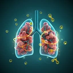
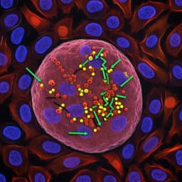
Omega-3 polyunsaturated fatty acids ameliorate PM2.5 exposure induced lung injury in mice through remodeling the gut microbiota and modulating the lung metabolism
J. Li, Y. Chen, et al.
A negative feedback loop between TET2 and leptin in adipocyte regulates body weight
Q. Zeng, J. Song, et al.
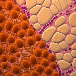
Maternal high-fat diet programs white and brown adipose tissue lipidome and transcriptome in offspring in a sex- and tissue-dependent manner in mice
C. Savva, L. A. Helguero, et al.


