
Medicine and HealthNature Communications
3D high-density microelectrode array with optical stimulation and drug delivery for investigating neural circuit dynamics
H. Shin, S. Jeong, et al.
Discover the groundbreaking research by Hyogeun Shin, Sohyeon Jeong, Ju-Hyun Lee, Woong Sun, Nakwon Choi, and Il-Joo Cho as they unveil a revolutionary 3D high-density multifunctional microelectrode array. This innovative device enhances our capability to monitor and modulate neural activities, paving the way for advanced in vitro studies in neural circuit dynamics.
Related Publications
Explore these studies to deepen your understanding
Adjacent work that informs or extends this paper's methodology and findings.
Medicine and Health
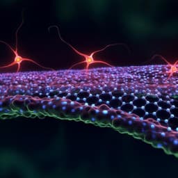
A conducting polymer-based array with multiplex sensing and drug delivery capabilities for smart bandages
L. Meng, S. Liu, et al.
Medicine and Health
Nanoporous graphene-based thin-film microelectrodes for in vivo high-resolution neural recording and stimulation
D. Viana, S. T. Walston, et al.

Engineering and Technology
3D-printing-assisted flexible pressure sensor with a concentric circle pattern and high sensitivity for health monitoring
J. Lee and H. So
Engineering and Technology
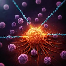
Nanoparticles and convergence of artificial intelligence for targeted drug delivery for cancer therapy: Current progress and challenges
R. P. Singh, A. Natarajan, et al.


