Medicine and Healthnpj Microgravity
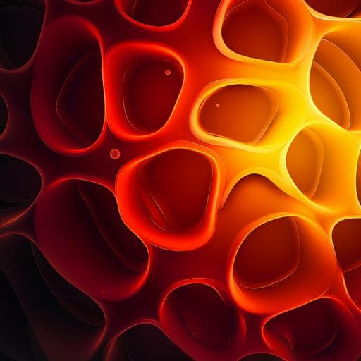
3D cell culture using a clinostat reproduces microgravity-induced skin changes
D. H. Choi, B. Jeon, et al.
Discover groundbreaking research on how microgravity influences skin health, revealing critical changes such as increased endothelial thickness and altered collagen expression. This study, conducted by Dong Hyun Choi, Byoungjun Jeon, Min Hyuk Lim, Dong Hun Lee, Sang-Kyu Ye, Seung-Yong Jeong, and Sungwan Kim, highlights the innovative use of a 3D clinostat to simulate microgravity's effects on skin physiology.
Related Publications
Explore these studies to deepen your understanding
Adjacent work that informs or extends this paper's methodology and findings.
Medicine and Health
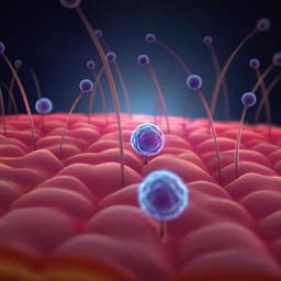
Single-cell transcriptomics reveals aberrant skin-resident cell populations and identifies fibroblasts as a determinant in rosacea
M. Chen, L. Yang, et al.
Medicine and Health
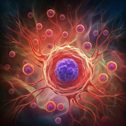
Engineering a niche supporting hematopoietic stem cell development using integrated single-cell transcriptomics
B. Hadland, B. Varnum-finney, et al.

Environmental Studies and Forestry
Marine protected areas do not prevent marine heatwave-induced fish community structure changes in a temperate transition zone
R. M. Freedman, J. A. Brown, et al.
Space Sciences
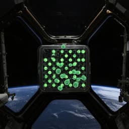
Surface tension enables induced pluripotent stem cell culture in commercially available hardware during spaceflight
M. Mozneb, M. Arzt, et al.


