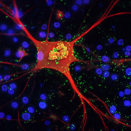
Introduction
Optical imaging, particularly near-infrared fluorescence imaging (NIRF), is widely used in preclinical studies, but suffers from limitations like autofluorescence, poor tissue penetration due to short excitation wavelengths, and excitation leakage. Chemiluminescence imaging, requiring no external excitation light, offers advantages in tissue penetration. However, its application is limited by low sensitivity and probe irreversibility. This research addresses these limitations by developing a turn-on chemiluminescence probe for in vivo imaging of amyloid-beta (Aβ) species, a hallmark of Alzheimer's disease. While numerous smart (turn-on) fluorescence probes exist for Aβ, turn-on chemiluminescence probes are rare. This study aims to design and validate a highly sensitive chemiluminescence probe to overcome the challenges of existing techniques and provide a powerful tool for Alzheimer's disease research and potential clinical applications.
Literature Review
The authors review existing chemiluminescence scaffolds, including dioxetane, luminol, and others, noting their limitations for in vivo imaging, especially in deep tissue like the brain. They discuss the challenges of existing fluorescence probes and highlight the scarcity of smart chemiluminescence probes for amyloid-beta detection. The research builds upon previous work by the same group on developing smart NIRF probes for Aβ, providing a context for the development of a chemiluminescence equivalent. The existing literature emphasizes the need for improved sensitivity and tissue penetration for in vivo amyloid imaging, driving the need for innovative approaches like the one presented in this paper.
Methodology
The researchers designed ADLumin-1, a chemiluminescence probe consisting of two moieties: moiety A (imidazo[1,2-a]pyrazin-3(7H)-one) for chemiluminescence and moiety B for binding to Aβ. The synthesis of ADLumin-1 is detailed. Spectral characterization showed an emission peak around 590 nm in DMSO and 540 nm in PBS with 10% DMSO. The authors investigated the probe's response to various reactive oxygen species (ROS), finding that oxygen dependence was the primary cause of chemiluminescence. A proposed mechanism for oxygen-dependent auto-oxidation is presented. The probe's binding to Aβ40 aggregates was confirmed through fluorescence spectroscopy (100-fold increase, Kd ≈ 2.1 µM) and molecular docking studies, which identified the probe's binding site in the Aβ fibril structure. In vitro chemiluminescence studies using Aβ aggregates in PBS buffer showed a 216-fold increase in signal. The researchers further demonstrated the probe's function in mouse brain homogenate (11.6-fold increase). The in vivo application of ADLumin-1 was investigated via brain slice incubation, two-photon imaging in live 5xFAD mice, ex vivo histology, and whole brain IVIS imaging. The study also explored the use of Dual-Amplification of signal via Chemiluminescence Resonance Energy Transfer (DAS-CRET) using ADLumin-1 as the donor and CRANAD-3 (a near-infrared fluorescence probe) as the acceptor, both non-conjugated. In vivo DAS-CRET imaging involved subcutaneous injection in nude mice to assess deep tissue penetration. In vivo whole-brain and eye imaging in 5xFAD and wild-type mice using DAS-CRET were performed using IVIS with open and specific filters, with spectral unmixing applied to enhance signal discrimination. Detailed protocols for oxygen and ROS sensitivity tests, Aβ40 aggregate preparation, fluorescence spectroscopy, in vitro chemiluminescence studies, binding affinity determination, in vitro and ex vivo histological studies, in vivo two-photon imaging, in vivo DAS-CRET mimic studies and in vivo chemiluminescence and DAS-CRET imaging are provided.
Key Findings
ADLumin-1, a novel turn-on chemiluminescence probe, demonstrated significant signal amplification in the presence of Aβ aggregates both in vitro (216-fold increase in PBS, 11.6-fold in brain homogenate) and in vivo (1.8-fold difference between 5xFAD and wild-type mice). Two-photon imaging revealed ADLumin-1's ability to cross the blood-brain barrier and effectively label both Aβ plaques and cerebral amyloid angiopathy (CAA) in vivo. High signal-to-noise ratios were observed for plaques and CAA (SNR≈17 and 26, respectively). The study showed that the large full width at half maximum (FWHM) of ADLumin-1's emission contributed to effective deep tissue penetration, comparable to firefly luciferin. DAS-CRET significantly amplified the chemiluminescence signal in vitro (133-fold increase in PBS with Aβ, 11.4-fold in brain homogenate) and in vivo (2.25-fold difference between 5xFAD and wild-type mice using 640 nm filter, and 3.22-fold after spectral unmixing), demonstrating the feasibility of this approach for enhancing chemiluminescence signal and extending emission into the near-infrared window. In vivo DAS-CRET also showed significant signal amplification in ocular imaging (2.11-fold difference between 5xFAD and wild-type mice). Higher Aβ levels were detected in the noses of 5xFAD mice compared to wild-type, a finding that requires further investigation.
Discussion
The findings demonstrate the successful development and validation of ADLumin-1, a highly sensitive and specific turn-on chemiluminescence probe for Aβ in vivo. The probe's ability to efficiently cross the blood-brain barrier and provide excellent contrast for Aβ plaques and CAA opens new avenues for Alzheimer's disease research. The successful implementation of DAS-CRET further enhances the sensitivity and deep tissue penetration capability of the imaging technique, providing a significant advantage over existing methods. The observation of increased Aβ levels in the noses of 5xFAD mice warrants further exploration as a potential non-invasive diagnostic marker. The potential extension of this technology to other aggregating-prone proteins highlights its broad applicability in neurodegenerative disease research.
Conclusion
This study successfully developed and validated ADLumin-1, a novel turn-on chemiluminescence probe for Aβ detection, and demonstrated the effectiveness of DAS-CRET for signal amplification and improved tissue penetration. This approach holds significant promise for preclinical research and potential translational applications in Alzheimer's disease diagnosis and monitoring of therapeutic efficacy. Future research could focus on exploring the potential of this technology for detecting Aβ in other tissues and applying it to other neurodegenerative diseases.
Limitations
While the study demonstrates promising results, limitations include the relatively short emission wavelength of ADLumin-1 (though this was addressed using DAS-CRET), the reliance on a transgenic mouse model, and the need for further validation in larger animal models and ultimately human patients. The proposed mechanism of auto-oxidation warrants further investigation to fully elucidate the underlying chemistry. The observation of higher Aβ signals in the nose requires further investigation to confirm its clinical significance.
Related Publications
Explore these studies to deepen your understanding of the subject.