
Food Science and Technologynpj Science of Food
Systematic evaluation of antimicrobial food preservatives on glucose metabolism and gut microbiota in healthy mice
P. Li, M. Li, et al.
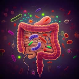
This study reveals the surprising impact of common antimicrobial preservatives on glucose metabolism and gut microbiota in healthy mice. Conducted by a team from the State Key Laboratory of Food Science and Technology, Nanchang University, the research highlights that even biogenic preservatives like nisin can lead to significant metabolic disruptions.
Related Publications
Explore these studies to deepen your understanding
Adjacent work that informs or extends this paper's methodology and findings.
Medicine and Health
Ablation of the gut microbiota alleviates high-methionine diet-induced hyperhomocysteinemia and glucose intolerance in mice
W. Li, Y. Jia, et al.

Biology
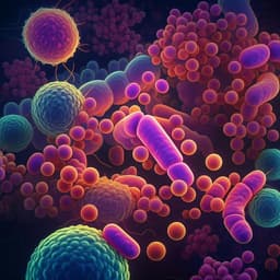
Gut microbiota determines the social behavior of mice and induces metabolic and inflammatory changes in their adipose tissue
O. Agranyomi, S. Meininger-mordechaï, et al.
Food Science and Technology
Effects of high fructose corn syrup on intestinal microbiota structure and obesity in mice
X. Wang, L. Zhu, et al.
Medicine and Health
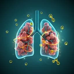
Omega-3 polyunsaturated fatty acids ameliorate PM2.5 exposure induced lung injury in mice through remodeling the gut microbiota and modulating the lung metabolism
J. Li, Y. Chen, et al.


