BiologyNature Plants
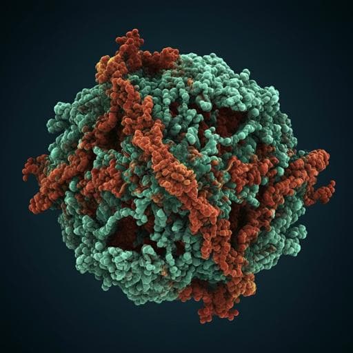
Structural basis for the activation of plant bunyavirus replication machinery and its dual-targeted inhibition by ribavirin
J. Li, L. Cao, et al.
This groundbreaking research by Jia Li, Lei Cao, and colleagues explores the intricate mechanics of the tomato spotted wilt orthotospovirus RNA-dependent RNA polymerase. Unveiling five unique cryo-EM structures, the study reveals how a flexible loop senses viral RNA and activates the polymerase, while ribavirin disrupts this essential binding, offering new pathways for antiviral pesticide development.
Related Publications
Explore these studies to deepen your understanding
Adjacent work that informs or extends this paper's methodology and findings.
Medicine and Health
Structural basis for modulation of human Nav1.3 by clinical drug and selective antagonist
X. Li, F. Xu, et al.

Agriculture
Transcriptome and genome sequencing elucidates the molecular basis for the high yield and good quality of the hybrid rice variety Chuanyou6203
J. Ren, F. Zhang, et al.

Food Science and Technology
Identification and quantitation of the actual active components in bamboo juice and its oral liquid by NMR and UPLC-Q-TOF-MS
Q. Gao, D. Wang, et al.

Medicine and Health
A multimodal deep learning approach for the prediction of cognitive decline and its effectiveness in clinical trials for Alzheimer’s disease
C. Wang, H. Tachimori, et al.


