Medicine and HealthNature Communications
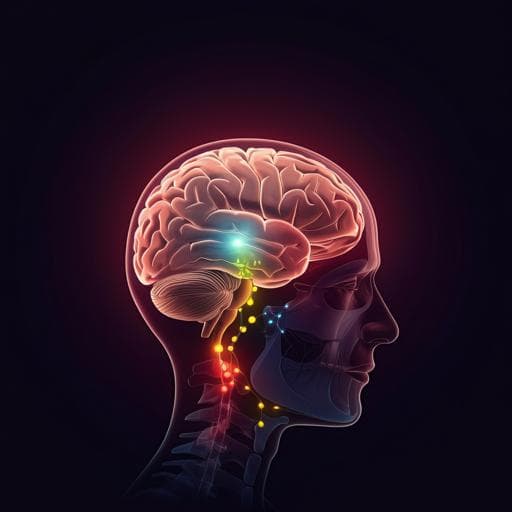
Stress-induced red nucleus attenuation induces anxiety-like behavior and lymph node CCL5 secretion
D. Shi, Y. Zhang, et al.
Explore groundbreaking research that reveals how stress influences immune responses through the brain-lymph node axis, led by researchers from the Shanghai Mental Health Center. Discover the connection between anxiety-like behavior and CCL5 levels, shedding light on the intricate interplay between the brain and immune system.
Related Publications
Explore these studies to deepen your understanding
Adjacent work that informs or extends this paper's methodology and findings.

Medicine and Health
Music prevents stress-induced depression and anxiety-like behavior in mice
Q. Fu, R. Qiu, et al.
Biology
Prefrontal Cortex-Specific Knockdown of Neurexin-1 in Rats Induces Anxiety-Like Behavior, Repetitive Behaviors, and Altered Social Interactions: A Proteomic Study
D. Wu, S. Zhang, et al.
Psychology
Repeated exposure with short-term behavioral stress resolves pre-existing stress-induced depressive-like behavior in mice
E. Lee, J. Park, et al.
Medicine and Health
Brain mitochondrial diversity and network organization predict anxiety-like behavior in male mice
A. M. Rosenberg, M. Saggar, et al.


