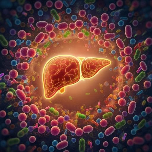
Stigmasterol attenuates hepatic steatosis in rats by strengthening the intestinal barrier and improving bile acid metabolism
Y. Zhang, Y. Gu, et al.
Explore these studies to deepen your understanding
Adjacent work that informs or extends this paper's methodology and findings.
Consumption of a high energy density diet triggers microbiota dysbiosis, hepatic lipidosis, and microglia activation in the nucleus of the solitary tract in rats
D. M. Minaya, A. Turlej, et al.

A diet high in sugar and fat influences neurotransmitter metabolism and then affects brain function by altering the gut microbiota
Y. Guo, X. Zhu, et al.
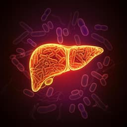
The interplay between dietary fatty acids and gut microbiota influences host metabolism and hepatic steatosis
M. Schoeler, S. Ellero-simatos, et al.
EGCG Attenuates CA1 Neuronal Death by Regulating GPx1, NF-κB S536 Phosphorylation and Mitochondrial Dynamics in the Rat Hippocampus following Status Epilepticus
A. V. Kozlov, S. Javadov, et al.


