logo
ResearchBunny Logo
Single-Cell Individualized Electroporation via Real-Time Impedance Monitoring

Medicine and Health

Single-Cell Individualized Electroporation via Real-Time Impedance Monitoring

Zhang

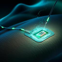
Explore cutting-edge research by Zhang, et al. on a revolutionary single-cell individualized electroporation method using a microelectrode array chip that enhances gene transfection efficiency while maintaining cell viability. This approach features real-time impedance monitoring and impressive results across various cell lines, paving the way for personalized electroporation techniques.

00:00
00:00
Playback language: English
Introduction
Electroporation (EP), a technique for introducing substances into cells by creating temporary pores in their membranes, suffers from drawbacks like substantial cell death due to inappropriate electric stimuli and incomplete membrane repair. Micro-electroporation (MEP) and nano-electroporation (NEP) techniques have been developed to improve precision and throughput, but challenges remain in real-time monitoring and cell viability optimization. Existing methods, like microfluidic channel EP, microcapillary EP, microarray EP, nanochannel EP, and others, have limitations in throughput, real-time monitoring, or invasiveness. Fluorescent dye methods can assess EP but require labeling and only show transient states. Patch-clamp is time-consuming and invasive. Electrical impedance spectroscopy (EIS) offers a label-free, real-time alternative. Microfluidic impedance cytometry (MIC) improves single-cell measurements but constrains cells. Electric cell-substrate impedance spectroscopy (ECIS) monitors adherent cells but lacks the single-cell resolution. This research addresses the need for a precise, high-throughput, and real-time monitoring system for optimized single-cell electroporation. The study aims to develop a novel method that balances perforation efficiency and cell viability by integrating cell positioning, MEP, and real-time impedance monitoring on a microelectrode array chip.
Literature Review
The literature extensively explores various micro- and nano-electroporation techniques. Microfluidic channel EP offers high throughput but lacks real-time response monitoring. Microcapillary EP allows selective EP but is slow. Microarray EP achieves high-throughput parallel transfection but struggles with real-time monitoring. NEP techniques focus the electric field on nanoscale portions of the cell membrane for precise delivery but also face challenges. Existing EP methods often result in substantial cell death due to uncontrolled electric stimuli and limited membrane repair. Current monitoring methods, such as fluorescence dye assays, have limitations in real-time monitoring and dynamic tracking. Electrical impedance spectroscopy (EIS) offers a label-free and real-time approach, and techniques like microfluidic impedance cytometry (MIC) and electric cell-substrate impedance spectroscopy (ECIS) have been employed, but each has its drawbacks in single-cell resolution, cell recovery, or throughput.
Methodology
The researchers developed a microelectrode array chip integrating cell positioning, MEP, and impedance monitoring. The system uses a function generator for electric signals, an impedance analyzer for real-time measurements, and a syringe pump for fluid control. The chip consists of sextupole-electrode units with center microelectrodes (7 × 7 µm²) for EP and impedance measurement, and quadrupole positioning electrodes (100 µm diameter, 100 µm gap) for cell manipulation via negative dielectrophoresis (nDEP). Cells are positioned using nDEP, then cultured for adhesion. EP is performed using electric pulses on the center electrodes, with impedance measured simultaneously. The center microelectrodes were modified with gold nanostructures to enhance measurement sensitivity. Experiments were conducted using HeLa, MCF-7, and 293T cells. EP parameters (pulse amplitude, number, width, frequency) were varied to assess their effects on impedance changes. Impedance indicators were defined: initial impedance, minimum impedance, recovery impedance, fall time, and recovery time. A comparative analysis using fluorescence (propidium iodide and Calcein-AM) was performed to validate the impedance indicators.
Key Findings
The study demonstrated successful single-cell positioning and electroporation using the microelectrode array chip. Real-time impedance monitoring accurately tracked the dynamic cell response to electroporation. The defined impedance indicators (initial descent impedance, recovery impedance, fall time, recovery time) effectively characterized perforation efficiency and cell viability. Initial descent impedance correlated with perforation efficiency, while recovery impedance correlated with cell viability. Increasing pulse amplitude, number, width, and frequency enhanced perforation but reduced viability. Optimal parameters were identified (pulse width of 100 µs and pulse frequency of 1 Hz). The fluorescence assay validated the impedance-based assessment of perforation efficiency and viability. The results demonstrate the feasibility of single-cell individualized EP optimization using real-time impedance monitoring. The method shows promise for diverse cell lines, suggesting potential in personalized therapy and diagnostics.
Discussion
The findings show that real-time impedance monitoring provides a powerful tool for optimizing single-cell electroporation. The established impedance indicators offer a label-free and quantitative method to evaluate both perforation efficiency and cell viability. The ability to dynamically adjust electroporation parameters based on real-time feedback allows for personalized treatment strategies, maximizing transfection success while minimizing cell damage. This approach offers significant advantages over traditional methods, paving the way for more effective gene therapy and drug delivery.
Conclusion
This research successfully demonstrated a single-cell individualized electroporation method using real-time impedance monitoring. The developed microelectrode array chip and the defined impedance indicators enable precise control and optimization of electroporation parameters, balancing perforation efficiency and cell viability across various cell lines. The method offers promising potential for personalized medicine and diagnostics. Future studies could investigate the application of this method in various therapeutic applications and explore advanced materials for further optimization of the chip's design and performance.
Limitations
The study primarily focused on three cell lines. Further investigations are needed to validate the method across a wider range of cell types and to assess its effectiveness with different types of delivery molecules. The current setup is optimized for in vitro studies. Adapting the method for in vivo applications presents a significant challenge that requires further development.
Listen, Learn & Level Up
Over 10,000 hours of research content in 25+ fields, available in 12+ languages.
No more digging through PDFs, just hit play and absorb the world's latest research in your language, on your time.
listen to research audio papers with researchbunny