
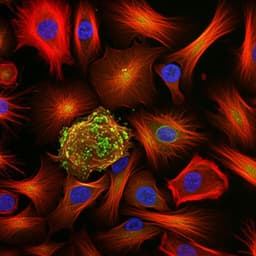
Single-cell guided prenatal derivation of primary fetal epithelial organoids from human amniotic and tracheal fluids
M. F. M. Gerli, G. Calà, et al.
Explore these studies to deepen your understanding
Adjacent work that informs or extends this paper's methodology and findings.

Single-cell guided prenatal derivation of primary fetal epithelial organoids from human amniotic and tracheal fluids
M. F. M. Gerli, G. Calà, et al.

Single-cell analysis of chromatin and expression reveals age- and sex-associated alterations in the human heart
D. F. Read, G. T. Booth, et al.
Single-cell RNA sequencing reveals the cellular and molecular heterogeneity of treatment-naïve primary osteosarcoma in dogs
D. T. Ammons, L. S. Hopkins, et al.
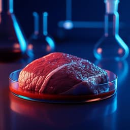
Cell-based, cell-cultured, cell-cultivated, cultured, or cultivated. What is the best name for meat, poultry, and seafood made directly from the cells of animals?
W. K. Hallman, W. K. H. Ii, et al.


