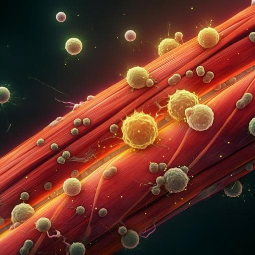
ROS-activated CXCR2+ neutrophils recruited by CXCL1 delay denervated skeletal muscle atrophy and undergo P53-mediated apoptosis
Y. Xiang, J. Dai, et al.
Explore these studies to deepen your understanding
Adjacent work that informs or extends this paper's methodology and findings.
Oral intake of rice overexpressing ubiquitin ligase inhibitory pentapeptide prevents atrophy in denervated skeletal muscle
R. Nakao, W. Shen, et al.

A tale of two paths to vaccine acceptance: self-interest and collective interest effect, mediated by institutional trust, and moderated by gender
O. Kol, D. Zimand-sheiner, et al.

Association Between Temporal Muscle Thickness and Skeletal Muscle Mass, Nutritional Status, and Physical Function in Patients With Post-stroke Hemiparesis: A Cross-Sectional Study
S. Terui, M. Maruyama, et al.
Disuse-associated loss of the protease LONP1 in muscle impairs mitochondrial function and causes reduced skeletal muscle mass and strength
Z. Xu, T. Fu, et al.


