Recent developments in multifunctional neural probes for simultaneous neural recording and modulation
H. Li, J. Wang, et al.
Explore these studies to deepen your understanding
Adjacent work that informs or extends this paper's methodology and findings.
Nanoporous graphene-based thin-film microelectrodes for in vivo high-resolution neural recording and stimulation
D. Viana, S. T. Walston, et al.
Fully bioresorbable hybrid opto-electronic neural implant system for simultaneous electrophysiological recording and optogenetic stimulation
M. Cho, J. Han, et al.
Using visible light to activate antiviral and antimicrobial properties of TiO2 nanoparticles in paints and coatings: focus on new developments for frequent-touch surfaces in hospitals
M. Schutte-smith, E. Erasmus, et al.
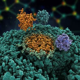
Tailored Functionalized Protein Nanocarriers for Cancer Therapy: Recent Developments and Prospects
R. J. Babu, A. K. Tiwari, et al.


