
Medicine and Health
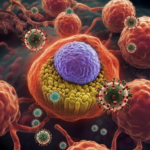
Prolonged SARS-CoV-2 RNA virus shedding and lymphopenia are hallmarks of COVID-19 in cancer patients with poor prognosis
A. Goubet, A. Dubuisson, et al.
This groundbreaking research reveals that cancer patients face increased severity of COVID-19 due to altered immune responses and prolonged viral RNA detection. The study's findings suggest a complex interplay between cancer therapies and COVID-19 immunopathology. Conducted by a team of expert researchers, these insights could be pivotal in developing strategies to reduce the cancer-related death risk associated with COVID-19.
~3 min • Beginner • English
Introduction
SARS-CoV-2 has caused a global pandemic of COVID-19. Patients with cancer appear more vulnerable to severe infection due to age, comorbidities, and therapy-related immunosuppression. Higher hospitalization and mortality rates have been reported in patients with malignancy, with hematological, lung, and breast cancers and metastatic disease conferring greater risk. Recent cancer treatments including surgery, chemotherapy, and immunotherapy independently worsen COVID-19 prognosis. The study explored independent cohorts of cancer patients diagnosed with COVID-19 to analyze host-viral dynamics (immunology, metabolism, metagenomics) and validated key findings across surges. The authors hypothesized that virus-induced lymphopenia, coinciding with T-cell exhaustion, metabolic pathway changes, and circulating bacterial DNA, is a poor prognostic factor in cancer, contributing to immunosuppression and chronic viral shedding.
Literature Review
Methodology
Design and cohorts: Multicohort observational study with prospective translational components. Cohorts included: Cancer_FR1_Translational Research (ONCOVID; NCT04341207) at Gustave Roussy (Discovery: Apr 10–May 4, 2020; Validation: May 5–Nov 25, 2020), healthcare workers (HCW) at Gustave Roussy, Cancer_FR2 case-control at IHU Méditerranée Infection (Marseille, France) with 175 cancer patients matched 1:2 to 350 cancer-free COVID-19 patients (Feb 27–Dec 15, 2020; >75% hydroxychloroquine and >96% azithromycin exposure), Canadian cancer cohort Cancer_CA (66 patients with longitudinal RT-qPCR), and Cancer_FR1_Clinical Routine (clinical records-based cohorts) plus Cancer_FR3 (Centre Léon Bérard, Lyon). Ethics approvals obtained as specified. Inclusion criteria in ONCOVID: adults with solid or hematologic malignancies under or recently treated by chemotherapy and/or immune checkpoint inhibitors; repeated SARS-CoV-2 RT-qPCR screening at hospital visits.
Definitions and outcomes: Duration of viral shedding = days from first positive to first negative RT-qPCR, requiring longitudinal monitoring and last positive to first negative interval <40 days to avoid overestimation. Long-term viral RNA shedding (LVS) defined as RT-qPCR positivity ≥40 days (median in Cancer_FR1_TR; ~95th percentile of HCW). Short-term viral RNA shedding (SVS) <40 days. COVID-19 severity categorized as mild, moderate (pneumonia on CT, hospitalization, oxygen <9 L/min), severe (ICU and/or oxygen >9 L/min). Absolute lymphocyte count (ALC) time-windows: PRE (−210 to −12 days before symptom onset), PER (−4 to +7 days around RT-qPCR diagnosis), POST (0 to 123 days after first negative RT-qPCR). Poor-prognosis strata combined Ct and ALC at diagnosis: Ct cutoff 25 (median; ~70% culture positivity threshold), ALC cutoff 800/mm³ (median among high viral load patients and CTCAE grade threshold).
Virology: Nasopharyngeal swabs collected; SARS-CoV-2 RT-qPCR performed (ELITech GeneFinder COVID-19 Plus RealAmp targeting RdRp, N, E on ELITe InGenius; or Applied Biosystems TaqPath COVID-19 CE-IVD targeting ORF1ab, N, S on QuantStudio 5). Ct values collected for Gustave Roussy assays. Subgenomic RNA RT-PCR (leader-E assay) per Wölfel et al. for selected samples.
Immunophenotyping and serology: High-dimensional spectral flow cytometry on whole blood assessing >80 markers of innate, B, and T cells. Panels quantified neutrophil maturation (CD101, CD10, CD16), monocyte subsets (CD14/CD16), activation/exhaustion markers (HLA-DR, CD38, PD-1, Eomes, TCF-1, Granzyme B, FasL, TOX), TFH (CXCR5, PD-1, ICOS, CD38), B-cell subsets (plasmablasts, transitional B, double-negative B). Serum analytes: multiplex 40-plex cytokines/chemokines, calprotectin, ultrasensitive IFN-α2a. Anti-SARS-CoV-2 IgA/IgM/IgG (including anti-S1 RBD) measured using Maverick Multi-Antigen Serology Panel.
Metabolomics: Serum metabolome by combined untargeted and targeted MS profiling >221 metabolites. Targeted pathways included bile acids (LC-MS/MS) and polyamines (LC-MS/MS). Additional profiling of SCFA and intracellular metabolites using UHPLC-Q-Exactive.
Metagenomics: Whole-blood 16S rDNA (V3-V4) sequencing from leukocyte-associated DNA; contamination-aware pipeline (FROGS), negative controls at extraction and PCR; taxonomy assignment via Silva database; LEfSe for differential taxa. Relative abundance assessed for families including Enterobacteriaceae and Micrococcaceae.
Clinical data: Demographics, comorbidities, ECOG PS, cancer type (solid vs hematologic), stage (localized/locally advanced/metastatic), treatment exposures and delays, hospitalization/ICU, deaths.
Statistics: Group comparisons via one-way ANOVA (Kenward-Roger), Mann-Whitney for two-group tests, Spearman correlations, hierarchical clustering, redundancy analysis (RDA) with permutation tests to relate biological variance to clinical covariates (virus shedding status, severity, age, malignancy type, metastatic status, diagnostic Ct). Kaplan–Meier for time to RT-qPCR negativity and overall survival. Cox regression (univariate and multivariate) for survival, stratified by cohort and adjusted for age, ECOG PS, gender, metastatic/hematological status. Significance P<0.05.
Key Findings
Viral kinetics and shedding:
- Cancer patients shed SARS-CoV-2 RNA significantly longer than healthcare workers (HCW): median 40 days (range 6–137) versus 21 days (range 7–53), log-rank P<0.0001; adjusted HR 2.88 (95% CI 1.42–5.85), P=0.00291 (Cancer_FR1_TR vs HCW).
- In FR2 case-control, RT-qPCR positivity length was longer in cancer vs cancer-free patients (median 8 vs 6 days), P=0.03; proportion with shedding >16 days (90th percentile of non-cancer) higher in cancer (P<0.0015).
- In Canada (Cancer_CA), 26% remained PCR-positive after 40 days.
- LVS (≥40 days) occurred more in hematologic malignancies than solid tumors (86% vs 43%, P=0.04, FR1_TR) and associated with advanced/metastatic disease (P=0.011), and with increased moderate COVID-19 severity in multiple cohorts (e.g., FR1_TR P=0.032; FR1_CR P=0.011).
- Lower diagnostic Ct values (higher viral load) correlated with longer shedding duration; Ct at diagnosis significantly anticorrelated with shedding duration; LVS patients had lower Ct at diagnosis than SVS across cohorts.
- RDA showed within 30 days from diagnosis, 18% variance in biological parameters explained by components influenced by shedding status (SVS vs negative P=0.037; LVS vs negative P=0.001), COVID-19 severity, age (P=0.0514), hematologic vs solid malignancy (P=0.001), metastatic status (P=0.0059), Ct at diagnosis (>25 vs <25, P=0.0738).
Immunological hallmarks:
- LVS patients displayed immunotypes typical of severe COVID-19 early after diagnosis: increased immature neutrophils (CD101+/− CD10+/− CD16−), decreased nonconventional monocytes (CD16+ CD14low/−) and decreased CD169+ HLA-DR+ conventional monocytes; generalized lymphopenia.
- Increased activated circulating TFH (CXCR5+ PD-1high CD4+ with ICOS and CD38) and plasmablasts (CD19low CD27hi CD38hi), reduced transitional and double-negative B cells; immature PMN and low double-negative B predicted LVS.
- Sustained elevation of type I IFN (IFN-α2a) in LVS versus SVS, controls, and recovered; IFN-α2a levels anticorrelated with anti-S1 RBD IgG titers.
- Expansion of non-naive CD8+ T cells expressing PD-1, Eomes, TCF-1, Granzyme B, and FasL, with features of exhaustion and apoptosis; frequencies positively correlated with duration of PCR positivity (e.g., PD-1+ non-naive CD8+ r≈0.68–0.78; P<0.01 to 0.0018 in reported panels).
Lymphopenia and prognosis:
- In cancer patients, ALC at diagnosis anticorrelated with duration of RT-qPCR positivity (r=−0.35, P=0.003). ALC dropped dramatically during acute COVID-19 in patients who later developed LVS, more than in SVS, and recovered post-clearance, indicating virus-induced lymphopenia. Higher viral load (Ct<25) associated with more pronounced lymphopenia.
- In cancer-free controls, no correlation between ALC and shedding duration (r=0.05, P=0.3); high viral load did not associate with lymphopenia at diagnosis or acute phase (P=0.11).
- Combined prognostic stratification (Discovery cohort, n=110; FR1+FR3): patients with ALC<800/mm³ and Ct<25 at diagnosis (21% of cohort) had dismal survival with ~40% deaths at 3 months; multivariate Cox: higher Ct reduced risk (HR 0.841 per unit, 95% CI 0.776–0.911; P=0.00002), higher ALC reduced risk (HR 0.282, 95% CI 0.119–0.672; P=0.0043); age (HR 1.042; P=0.0043) and ECOG>2 (HR 4.547; P=0.0001) increased risk; metastatic/hematologic status HR 2.747; P=0.0322.
- Validation cohort (n=116; FR1+FR3): ALC<800 & Ct<25 group (32.7%) again had the poorest survival (>40% deaths by day 50).
Metabolomics and metagenomics correlates:
- Serum metabolomics differentiated LVS from SVS; secondary bile acids (murideoxycholic acid, deoxycholic acid, hyodeoxycholic acid, ursodeoxycholic acid) were decreased in LVS versus SVS/controls and correlated with lower ALC; DOC also associated with increased disease severity in noncancer patients.
- Polyamine pathway: N1,N8-diacetylspermidine increased in LVS (and in severe noncancer COVID-19) and negatively correlated with ALC, linking to lymphocyte fitness decline.
- Blood 16S rDNA: Enterobacteriaceae DNA overrepresented in LVS; its abundance anticorrelated with serum CCL22 and correlated positively with exhausted CD8+ T-cell markers. Trend toward increased Micrococcaceae in LVS and enrichment in poor-prognosis (ALC<0.8 & Ct<25) group.
Discussion
The study demonstrates that cancer patients experience prolonged SARS-CoV-2 RNA shedding, linked to higher initial viral loads and immunopathologic signatures characteristic of severe COVID-19. Prolonged shedding (LVS) associates with lymphopenia and expansion of exhausted CD8+ T cells, sustained type I IFN responses, increased plasmablasts and activated TFH, and myeloid dysregulation. Virus-induced lymphocyte loss, superimposed on cancer- and therapy-related immunosuppression, compromises viral clearance and predicts early mortality. Metabolic alterations—depletion of secondary bile acids and accumulation of diacetylspermidine—together with increased circulating leukocyte-associated bacterial DNA (Enterobacteriaceae, Micrococcaceae) suggest gut permeability/dysbiosis and disrupted lymphocyte maintenance pathways contribute to lymphopenia and T-cell exhaustion. These mechanisms may sustain a positive feedback loop of interferon-stimulated gene expression (including ACE2) and persistent inflammation, hindering virus clearance and potentially impairing antitumor immunity. Clinically, combining Ct and ALC at diagnosis stratifies risk, identifying patients with markedly worse survival across two pandemic surges. The findings argue for careful management of immunosuppressive therapies during acute infection and support strategies that both neutralize virus and restore lymphopoiesis.
Conclusion
Cancer patients with COVID-19 frequently exhibit prolonged SARS-CoV-2 RNA shedding that correlates with high initial viral loads, lymphopenia, T-cell exhaustion, and specific metabolic and metagenomic signatures. The combination of low ALC (<800/mm³) and high viral load (Ct<25) at diagnosis robustly predicts poor short-term survival and was validated across independent cohorts and surges. The work highlights virus-induced lymphocyte loss as a key driver of poor outcomes in cancer and suggests that preventing or reversing COVID-19-related lymphopenia—potentially via passive immunization and agents promoting lymphopoiesis (e.g., IL-7, G-CSF)—alongside patient stratification by Ct, ALC, and shedding duration, may improve prognosis and enable safer cancer therapy. Future research should test targeted interventions to restore immune competence, dissect causal links between bile acid and polyamine metabolism and lymphocyte fitness, and clarify the role of bacterial translocation in sustaining immunopathology.
Limitations
Prolonged RT-qPCR positivity reflects viral RNA detection and may not confirm ongoing infectiousness; although subgenomic RNA assays were used in part, replication competence was not universally established. Cohorts were heterogeneous across centers and countries with variable testing platforms and treatment exposures (e.g., hydroxychloroquine/azithromycin in FR2), which may confound shedding duration. Some analyses involved limited sample sizes (e.g., flow cytometry, metabolomics, blood microbiome), with missing Ct/ALC data for subsets. Observational design precludes causal inference regarding metabolic and metagenomic associations with lymphopenia. Variability in timing of sampling relative to symptom onset could affect immunologic and metabolic measurements.
Related Publications
Explore these studies to deepen your understanding of the subject.