Medicine and HealthNot specified in provided text
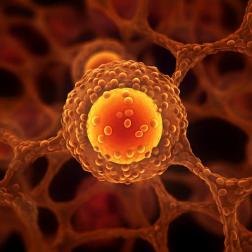
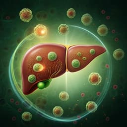
Perilipin 5 regulates hepatic stellate cell activation and high-fat diet-induced non-alcoholic fatty liver disease
X. Yin, L. Dong, et al.
Unlock the secrets of nonalcoholic fatty liver disease (NAFLD) with groundbreaking research by Xuecui Yin and colleagues. This study reveals how Perilipin 5 influences hepatic stellate cell activation, impacting lipid homeostasis, glucose regulation, and mitochondrial health. Discover the potential of PLIN5 in combating liver fibrosis and enhancing metabolic performance!
Related Publications
Explore these studies to deepen your understanding
Adjacent work that informs or extends this paper's methodology and findings.
Medicine and Health
Alantolactone attenuates high-fat diet-induced inflammation and oxidative stress in non-alcoholic fatty liver disease
J. Wang, Y. Jiang, et al.
Medicine and Health
Potential role of inflammation in relation to dietary sodium and β-carotene with non-alcoholic fatty liver disease: a mediation analysis
Y. Chen, M. Wu, et al.

Medicine and Health
Peripancreatic adipose tissue protects against high-fat-diet-induced hepatic steatosis and insulin resistance in mice
B. Chanclón, Y. Wu, et al.
Medicine and Health
Pyruvate dehydrogenase kinase 1 and 2 deficiency reduces high-fat diet-induced hypertrophic obesity and inhibits the differentiation of preadipocytes into mature adipocytes
H. Kang, B. Min, et al.


