Medicine and HealthBiomedicines
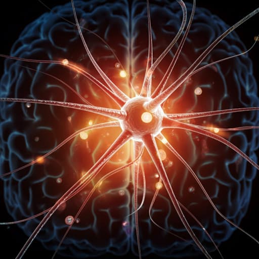
Neuroprotective Effects of Human-Induced Pluripotent Stem Cell-Derived Mesenchymal Stem Cell Extracellular Vesicles in Ischemic Stroke Models
G. Lu, X. Su, et al.
Discover the groundbreaking research by Gang Lu, Xianwei Su, Lihon Wang, Chi-Kwan Leu, Jingye Zhou, Zhiqiang Xiong, Wuming Wang, Hongbin Liu, and Wai-Yee Chan on the neuroprotective effects of extracellular vesicles derived from hiPS-MSCs in ischemic stroke. Their study reveals the potential of these EVs to reduce injury and promote recovery, paving the way for innovative cell-free therapies in stroke treatment.
Related Publications
Explore these studies to deepen your understanding
Adjacent work that informs or extends this paper's methodology and findings.
Medicine and Health
Two-year safety outcomes of iPS cell-derived mesenchymal stromal cells in acute steroid-resistant graft-versus-host disease
K. Kelly, A. J. C. Bloor, et al.

Medicine and Health
A framework for human evaluation of large language models in healthcare derived from literature review
T. Y. C. Tam, S. Sivarajkumar, et al.
Biology
Transplanted human iPSC-derived vascular endothelial cells promote functional recovery by recruitment of regulatory T cells to ischemic white matter in the brain
B. Xu, H. Shimauchi-ohtaki, et al.
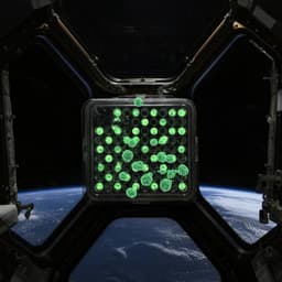
Space Sciences
Surface tension enables induced pluripotent stem cell culture in commercially available hardware during spaceflight
M. Mozneb, M. Arzt, et al.


