Medicine and HealthNature Nanotechnology
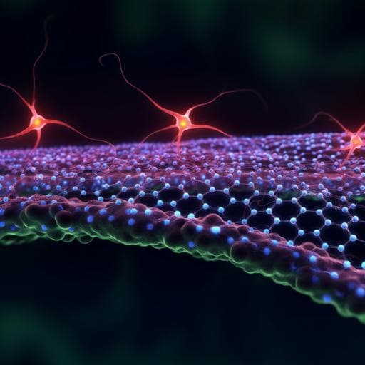
Nanoporous graphene-based thin-film microelectrodes for in vivo high-resolution neural recording and stimulation
D. Viana, S. T. Walston, et al.
Discover cutting-edge advancements in flexible neural interfaces with our innovative nanoporous graphene-based technology. This groundbreaking research led by a team from the Catalan Institute of Nanoscience and Nanotechnology and other esteemed institutions demonstrates exceptional recording fidelity and biocompatibility in chronic studies.
Related Publications
Explore these studies to deepen your understanding
Adjacent work that informs or extends this paper's methodology and findings.
Biology
Recent developments in multifunctional neural probes for simultaneous neural recording and modulation
H. Li, J. Wang, et al.

Engineering and Technology
High-resolution flexible iontronic skins for both negative and positive pressure measurement in room temperature wind tunnel applications
J. Wang, X. Wei, et al.

Biology
In vivo volumetric imaging of calcium and glutamate activity at synapses with high spatiotemporal resolution
W. Chen, R. G. Natan, et al.

Engineering and Technology
High-Q cavity interface for color centers in thin film diamond
S. W. Ding, M. Haas, et al.


