BiologyNature Communications
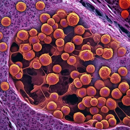
Multiplexed histology analyses for the phenotypic and spatial characterization of human innate lymphoid cells
A. Pascual-reguant, R. Köhler, et al.
This study by Anna Pascual-Reguant and colleagues unveils the intricate world of innate lymphoid cells (ILCs), exploring their localization and interactions within the tonsil microenvironment. By identifying IRF4 as a specific marker for tonsillar ILC3s, the researchers reveal shared niches with plasma cells, paving the way for deeper understanding of ILC biology through advanced histological techniques.
Related Publications
Explore these studies to deepen your understanding
Adjacent work that informs or extends this paper's methodology and findings.
Biology
Revised Estimates for the Number of Human and Bacteria Cells in the Body
R. Sender, S. Fuchs, et al.

Health and Fitness
Baseline imbalance and heterogeneity are present in meta-analyses of randomized clinical trials examining the effects of exercise and medicines for blood pressure management
M. A. Wewege, H. J. Hansford, et al.

Economics
Towards a healthier future for the achievement of SDGs: unveiling the effects of agricultural financing, energy poverty, human capital, and corruption on malnutrition
C. Ding, K. A. Khan, et al.

Medicine and Health
Generation and characterization of cardiac valve endothelial-like cells from human pluripotent stem cells
L. Cheng, M. Xie, et al.


